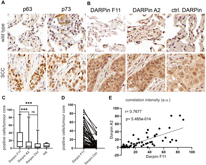
Fig. 6. DARPins A2 and F11 detect the p632/p732 hetero-tetramer in human NSCLC-SCC.
A Representative immunohistology stainings against endogenous p63 (clone 4A4) in matched non-transformed and human NSCLC-SCC. Scale bar = 1 mm or 2 mm, respectively. B Immunohistology analysis of non-transformed and matched NSCLC-SCC tumors exposed to HA-tagged control-DARPin and DARPins A2 and F11 as indicated, followed by HA-epitope specific antibodies. The tumor area is highlighted by dashed lines. Scale bar = 50 µm. C Quantitative analysis of IHC intensity in human SCC using DARPins F11 and A2, compared to control DARPin in mirco-array sections. The analysis was conducted with the image analysis software QuPath (0.4.2). D Parallel coordinate plots of total positive cells for DARPin F11 and control DARPin. Every connected dot represents a tumor sample of one patient with positive immunohistology signal in cells [%] of DARPin F11 or control DARPin. Mann-Whitney Test p > 0.001. E Pearson correlation between positive immunohistology signals of DARPin F11 versus DARPin A2 in human SCC tumor cores of a NSCLC tissue micro array (R = Pearson correlation coefficient, p = two tailed t test).

