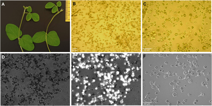
Figure 1.
Isolation of protoplasts from trifoliate leaves of soybean plantlets. (A) Fifteen-day-old plants showing trifoliate leaves of suitable size. (B, C) Protoplasts of freshly extracted (B) and purified cells (C) under the Motic AE2000 inverted microscope with × 20 and × 40 objectives, respectively. Black scale bar, 30 µm. (D, E) The protoplast viability was assessed by FDA staining and observed under both bright field (D) and fluorescence channel, and simultaneously merged images are depicted (E) using Axio Vert.A1 inverted microscope with a × 20 objective. (F) Division of protoplasts (shown by white arrows) at 4 days after isolation in culture medium. FDA, fluorescein diacetate.

