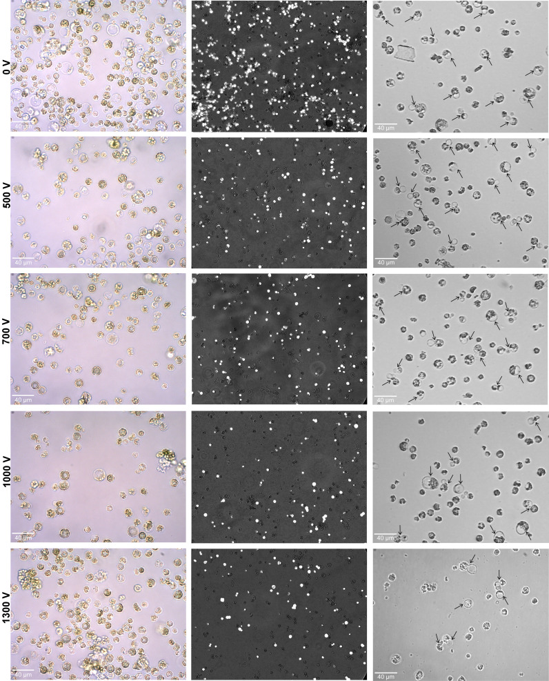
Figure 2.
Effect of various pulsing voltage on the protoplasts morphology, viability, and cell division efficiency following electro-transfection. Left panel: representative images of electroporated protoplasts after 24 h are shown. Middle panel: the merged fluorescence images (under bright and fluorescence field using Axio Vert.A1 inverted microscope with a × 20 objective) showing the FDA-stained viable cells after 24 h of electroporation. Right panel: images showing primary divisions (shown by black arrows) of electroporated protoplasts at 4 days after culture initiation. FDA, fluorescein diacetate.

