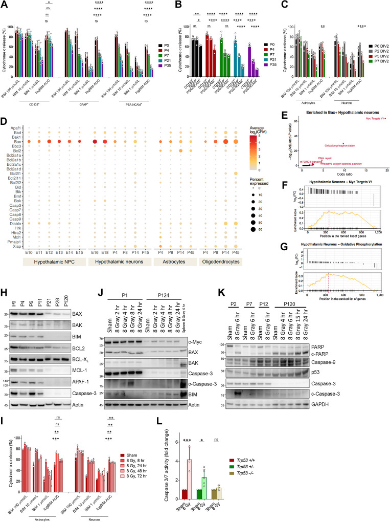
Figure 3.
Mechanism of xRT-induced NI. A, BH3 profiling of neural stem cells (CD133+), astrocytes (GFAP+), and neurons (PSA-NCAM+) isolated from mice of indicated ages. B, Comparison of priming in indicated cell types isolated from brain tissue from P5 animal. C, BH3 profiling of astrocytes and neurons cultured from neural stem cells collected at indicated ages. D, Expression of mitochondrial apoptosis-associated genes in indicated cell types from mid embryogenesis to adulthood. E, Gene set enrichment analysis on BAX-expressing hypothalamic neurons. F and G, Enrichment plots for Myc target genes and genes associated with oxidative phosphorylation in BAX-expressing neurons. H, Immunoblotting for BCL2 family proteins in brain tissue isolated from animals of indicated ages. Relative molecular weight markers are shown on left for all immunoblots. I, BH3 profiling of in vitro cultured astrocytes and neurons treated with 8 Gy ionizing radiation for the indicated time period. J, Immunoblotting for BCL2 family proteins in neonatal (P1) or adult (P124) brain tissue after 8 Gy IR treatment in vivo for indicated time period. K, Immunoblotting for markers of apoptosis in brain tissue collected from animals irradiated at indicated age for indicated time period. L, Caspase-3/7 glow assay measuring caspase-3/7 activity in brain tissue collected from animals of indicated genotype 5 hours after 8 Gy xRT treatment. Unless otherwise specified, comparison between groups was conducted by one-way or two-way ANOVA followed by Holm-Sidak post hoc multiple comparisons test. *, P < 0.05; **, P < 0.01; ***, P < 0.001; ****, P < 0.0001; ns, nonsignificant.

