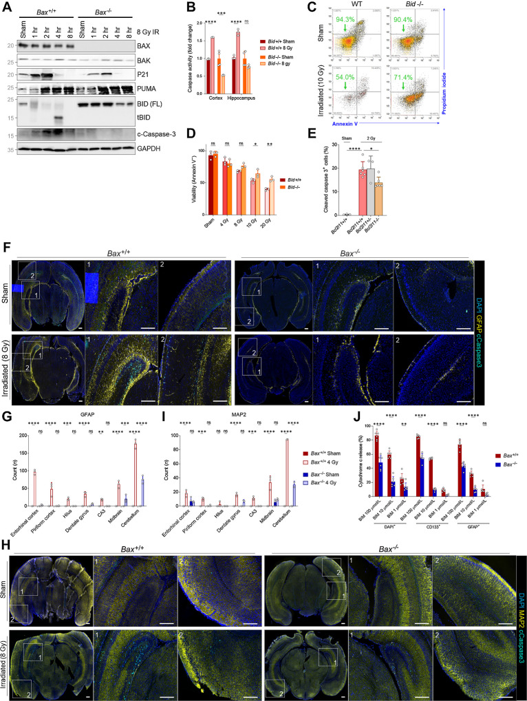
Figure 4.
BAX ablation protects from xRT-induced apoptosis in neural stem and progenitor cells. A, Immunoblotting for BCL2 family proteins in Bax+/+ or Bax−/− P1 animals at indicated time period after 8 Gy xRT. B, Caspase-3/7 glow assay measuring caspase-3/7 activity in Bid+/+ and Bid−/− animals in cortex and hippocampus regions of brain 5 hours after 8 Gy xRT. C, Representative flow cytometry plots of viability analysis of MEFs of indicated genotype 48 hours after irradiation. D, Summary of viability analysis performed in MEFs of indicated genotype 48 hours after irradiation with indicated doses. E, BIM contributes toward the radiation sensitivity of CGNPs, as Bim−/− mice have decreased numbers of apoptotic cC3+ cells following radiation treatment (2 Gy, 3 hours after treatment) when compared with controls. F, Representative images of whole-brain sections immunostained for indicated markers (DAPI, GFAP, cleaved caspase-3). Scale bars, 200 μm. G, Quantification of cells positive for cleaved caspase-3 and GFAP in indicated brain regions. H, Representative images of whole-brain sections immunostained for indicated markers. Scale bars, 200 μm. I, Quantification of cells positive for cleaved caspase-3 and MAP2 in indicated brain regions. Values presented are means + SD from two to three separate experiments. All groups were compared with two-way ANOVA with Holm-Sidak adjustment. J, BH3 profiling of P2 mouse brain tissue from Bax+/+ and Bax−/− animals with stains for nucleated cells (DAPI+), neural stem cells (CD133+), and astrocytes (GFAP+). Unless otherwise specified, comparison between groups was conducted by one-way or two-way ANOVA followed by Holm-Sidak post hoc multiple comparisons test. *, P < 0.05; **, P < 0.01; ***, P < 0.001; ****, P < 0.0001; ns, nonsignificant.

