Figure 5.
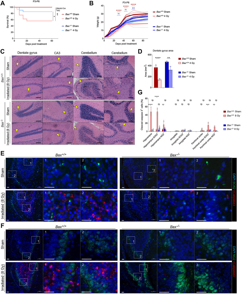
Knockout of BAX reduces xRT-induced mortality and neurodegeneration. A, Overall survival of mice of indicated genotypes irradiated at P3-P6. B, Weight measurements of mice of indicated genotypes irradiated at P3-P6. C, Histology of indicated brain regions from Bax+/+ versus Bax−/− animals 180 days after irradiation or sham treatment. Arrows, areas of reduced or retained cellularity. Scale bars, 100 μm. D, DG areas of mouse brains 8 to 9 months after indicated treatment. E and F, Representative images of brain sections from hippocampus immunostained with DAPI and antibodies for cleaved caspase-3 as well as SOX2 (E) and NG2 (F). Scale bars, 25 μm. G, Quantification of cells positive for cleaved caspase-3 and SOX2 or NG2 in indicated brain regions in Bax+/+ versus Bax−/− animals after irradiation or sham treatment. Unless otherwise specified, comparison between groups was conducted by one-way or two-way ANOVA followed by Holm-Sidak post hoc multiple comparisons test. *, P < 0.05; **, P < 0.01; ***, P < 0.001; ****, P < 0.0001; ns, nonsignificant.

