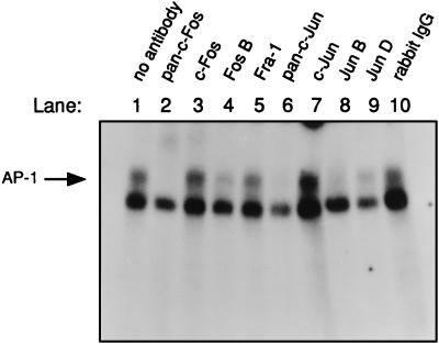
FIG. 4.
The superinduced AP-1 complex in ciprofloxacin-treated CD4+ T cells contained proteins from the c-Fos and c-Jun families. Cell extract from the CD4+ T cells exposed to ciprofloxacin for 3 h displayed in Fig. 3A was examined in supershifts. The upper band shows the specific AP-1 binding (see, for example, lanes 1 and 3). The lower band shows the unspecific binding to the AP-1 oligonucleotide sometimes observed. Rabbit polyclonal antibodies were preincubated with the extracts for 30 min on ice prior to addition of the AP-1 oligonucleotide. Unspecific rabbit immunoglobulins, which were used as a control serum, are indicated by rabbit immunoglobulin G (IgG) in lane 10. The free probe (shown in Fig. 3A) was run out from the gel. Similar results were obtained with the 2-h extract from control T cells shown in Fig. 3A (data not presented). The results are representative for two different blood donors.

