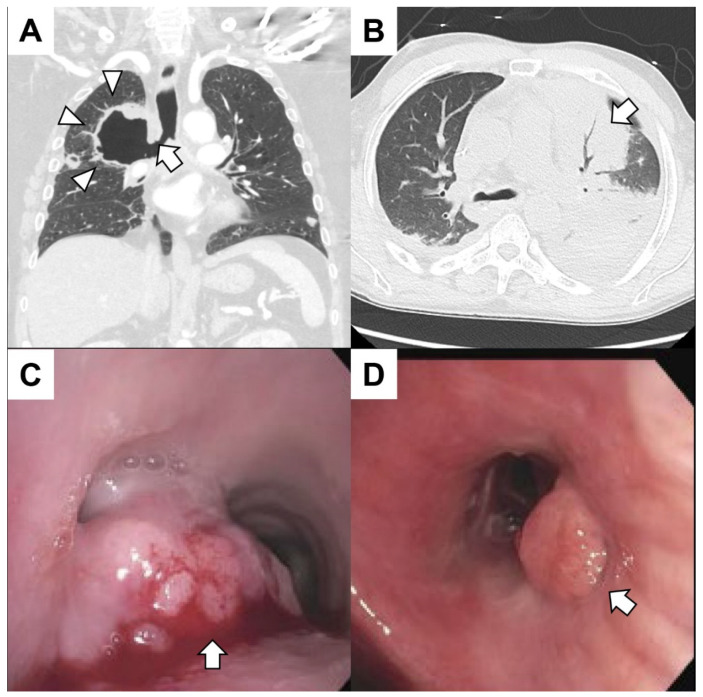
Figure 2.
(A) A patient with metastatic lung cancer treated with radiation 1 month prior to the right upper lobe presented with hemoptysis. The coronal CT of the chest revealed a cavitary lesion (arrowheads) contiguous with the right mainstem bronchus (arrow). The patient expired from massive hemoptysis shortly after presenting to the emergency center. (B) A patient with leukemia with pancytopenia (white blood cells 0.0 × 10⁹/L platelets 14,000 × 109/L) and fever presented with hemoptysis. The patient had sepsis with bacteremia. A chest CT revealed consolidation in the left upper lobe (arrow) with air bronchograms. (C) A patient with gastroesophageal junction cancer presented with dyspnea and mild hemoptysis. Bronchoscopy revealed a mass at the main carina obstructing 90% of the left mainstem and 20% of the right mainstem. The mass (arrow) was large, exophytic and friable. The tumor was debulked using rigid bronchoscopy, and argon plasma coagulation was used to achieve hemostasis. Biopsies were consistent with metastatic disease. (D) A patient with metastatic colorectal cancer had dyspnea and mild hemoptysis. The patient had mucopurulent and blood-tinged secretions emanating from the right middle lobe. An endoluminal tumor mass (arrow) obstructing the superior segment of the left lower lobe was found. The hemoptysis resolved after treatment of a respiratory infection.

