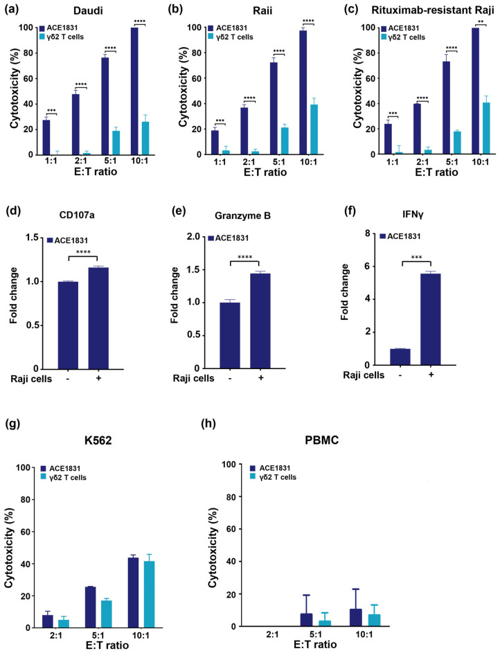
Figure 2.
Rituximab conjugation confers γδ2 T cells with superior cytotoxicity against CD20-expressing cancer cells. (a–c) ACE1831 and γδ2 T cells were co-incubated with CD20-expressing cancer cells. (a) Daudi, (b) Raji and (c) rituximab-resistant Raji cells at effector to target (E:T) ratios of 1:1, 2:1, 5:1 and 10:1, analyzed via CellTiter-Glo® luminescent cell viability assay after 4 h of co-incubation. (d) CD107a, (e) granzyme B and (f) IFNγ of ACE1831 in the absence and presence of Raji cells at an E:T ratio of 2:1 after 2 h of incubation were analyzed via flow cytometry. Each group was performed in triplicate from two donor lots, and the representative results are shown. Statistical analysis was performed using the t test. **, p < 0.01; ***, p < 0.001; ****, p < 0.0001. (g,h) ACE1831 and γδ2 T cells were co-incubated with (g) CD20-negative K562 and (h) donor PBMC cells at effector to target (E:T) ratios of 2:1, 5:1 and 10:1 and analyzed using a CellTiter-Glo® luminescent cell viability assay after 4 h of co-incubation.

