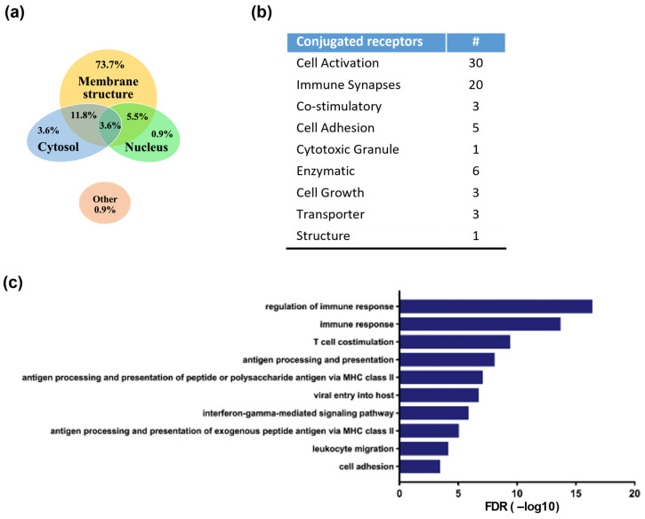
Figure 4.
Identification and functional analysis of rituximab-conjugated proteins using mass spectrometry. Rituximab-linked proteins of ACE1831 were immunoprecipitated and analyzed using mass spectrometry. Protein candidates with ACE1831/un-conjugated γδ2 T cell ratios larger than 550 and an average frequency less than 0.3 were identified as rituximab-linked proteins. The cellular component, biological process, KEGG pathway GO term and p-value were analyzed with the GO Consortium and the Database for Annotation, Visualization, and Integrated Discovery. (a) Proportion of identified rituximab-linked proteins in the cellular localization. (b) Functional categorization of rituximab-linked proteins. “#” stands for the number of conjugated receptors in each functional category. (c) Top 10 categories of biological processes among the identified rituximab-linked proteins.

