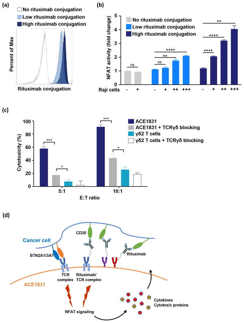
Figure 5.
T cell activation and cytotoxicity mediated by the antigen recognition of ACC-linked antibody. (a) Jurkat-NFAT-Luc cells were conjugated with different amounts of rituximab using ACC technology and were stained with anti-F(ab’)2 antibody to examine the levels of rituximab conjugated on Jurkat-NFAT-Luc cells. Un-conjugated Jurkat-NFAT-Luc cells (grey line) represent negative staining, and Jurkat-NFAT-Luc cells with low (light blue) and high (dark blue) rituximab conjugation are shown. Percent of Max is the highest point of each peak of the overlaid histogram. (b) Jurkat-NFAT-Luc cells conjugated with different amounts of rituximab were co-incubated with different Raji cell numbers (+, 5 × 10⁴; ++, 2 × 10⁵; +++, 5 × 10⁵), and NFAT signaling activation was determined based on NFAT-regulated luciferase activity. Each condition was applied in triplicate in two different experiments, and the representative results are shown. Mean ± SD. Statistical analysis was performed using a t test. **, p < 0.01; ****, p < 0.0001. (c) The effector cells were preincubated with or without 1 μg/mL of TCRγδ blocking antibody for 1 h at 37 °C. After 4 h of co-incubation with Raji cells, cytotoxicity against Raji cells was analyzed using a CellTiter-Glo® luminescent cell viability assay. Each condition was applied in triplicate in two different experiments, and the representative results are shown. Mean ± SD, *, p < 0.05, ***, p < 0.001. (d) Illustration delineating the activation of ACE1831 upon encountering CD20-expressing cancer cells.

