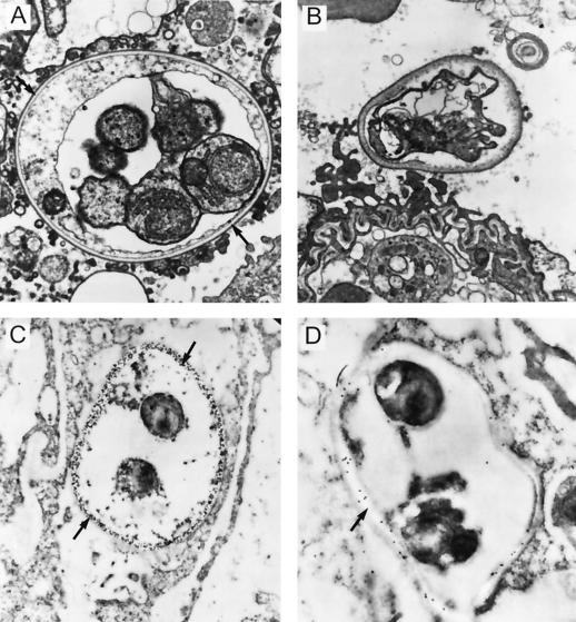
FIG. 7.
(A) Mature P. carinii cyst containing trophozoites in rat lung tissue. The electron-lucent layer of the cyst wall is the proposed location of β-1,3-glucan. (B) Cyst from a rat with acute PCP treated for 2 days with the pneumocandin L-733,560. The electron-lucent layer is absent, while the electron-dense layer has thickened and the wall has lost its rigid shape. (C) Section of the lung sample in panel A fixed for immunogold electron microscopy. The section was incubated with antiserum specific for β-1,3-glucan and labelled with immunogold. The β-1,3-glucan antibody reacts only with the translucent layer of the cyst wall. There is no reactivity seen with the surrounding lung tissue or with the trophozoites. (D) Section of the lung sample shown in panel B. The section was incubated with antibody made against L-733,560 and then labelled with immunogold. The antibody specifically targets what is left of the translucent layer, demonstrating that the pneumocandins localize in the cyst wall upon treatment of the animal.

