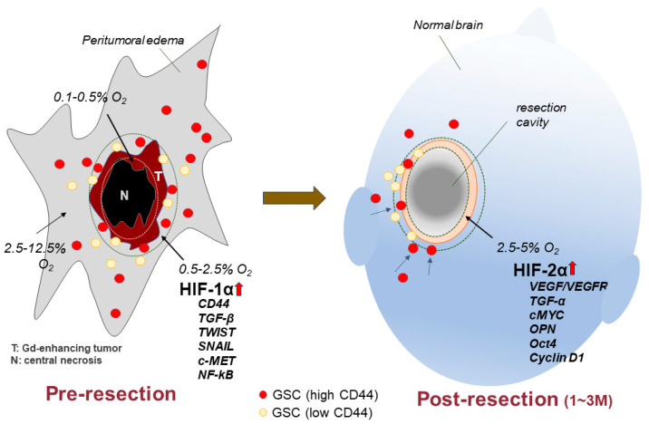
Figure 6.
Illustration of hypothetical differential expression of genes related to cell invasion/migration and cell proliferation in glioblastoma according to hypoxic conditions before tumor resection and 1–3 months (in the period of tissue repair) after tumor resection. HIF-1α is upregulated by severe hypoxia of 0.5–2.5% O2, corresponding to the tumor rim (tumor border area) before resection. In this area, HIF-1α target genes (including CD44, TGF-β, TWIST, SNAIL, cMET, and NF-kB) are activated, resulting in promotion of cell migration and invasion and enhancement of stemness. HIF-2α is upregulated by moderate hypoxia of 2.5–5% O2, corresponding to the marginal area of the resection cavity once tissue injury is repaired and presenting with moderate hypoxia. These cellular processes represent the phenotypic transition of GBM.

