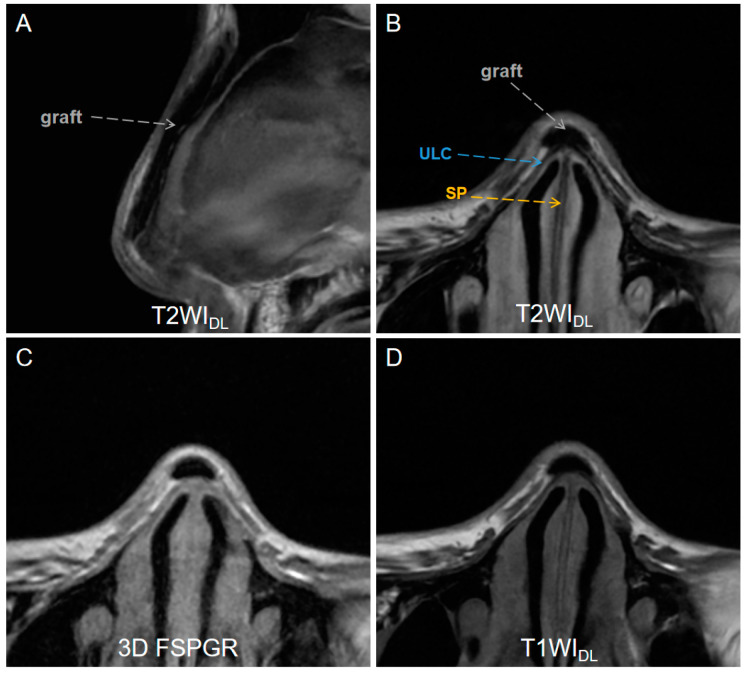
Figure 2.
A low-signal-intensity graft (gray dashed arrows) on (A) sagittal T2WIDL image (DLR T2-weighted FSE images), (B) axial T2WIDL image, (C) axial 3D FSPGR image (three-dimensional fast spoiled gradient-recalled images), (D) T2WIO (original T2-weighted FSE images), and (D) axial T1WIDL (deep learning–reconstructed T1-weighted FSE images) image of a 26-year-old female who underwent an MR examination one year after receiving augmentation rhinoplasty with autogenous ear cartilage. T2WIDL shows a clearer boundary between the graft and the tissue. DLR = deep-learning-based reconstruction, SP = septal cartilage (yellow dashed arrow), ULC = upper lateral cartilage (blue dashed arrow).

