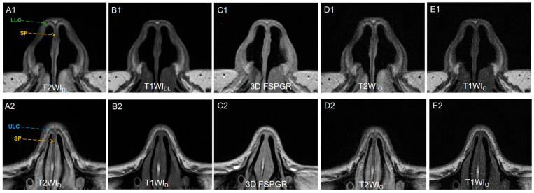
Figure 3.
(A1,A2) T2WIDL (DLR T2-weighted FSE images), (B1,B2) T1WIDL (DLR T1-weighted FSE images), (C1,C2) 3D FSPGR (three-dimensional fast spoiled gradient-recalled images), (D1,D2) T2WIO (original T2-weighted FSE images), and (E1,E2) T1WIO (original T1-weighted FSE images) in axial view of a 25-year-old female nasal cartilage. The overall image quality and contrast of FSEDL images showed better than any other image sets because of less noise. T2WIDL showed the best anatomical structure of nasal cartilage for higher contrast. No significantly different display of ULC between T1WIDL and 3D FSPGR was observed (p > 0.05) while 3D FSPGR showed relatively poor image quality of SP. DLR = deep-learning-based reconstruction, SP = septal cartilage (yellow dashed arrow), LLC = lower lateral cartilage (green dashed arrow) and ULC = upper lateral cartilage (blue dashed arrow).

