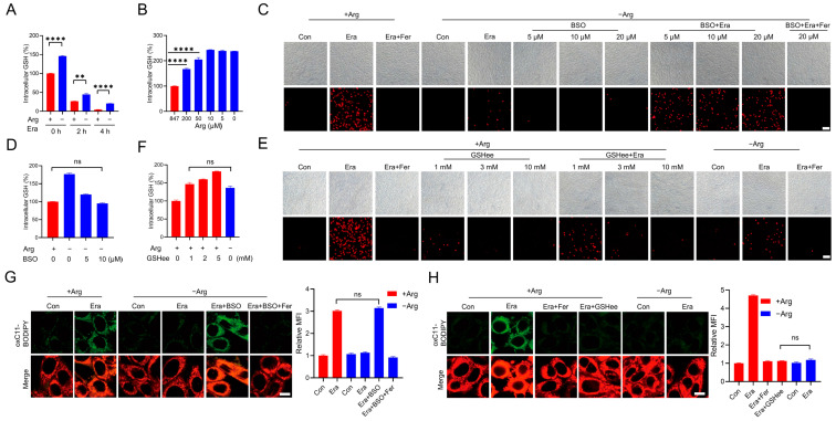
Figure 2.
Arginine promotes erastin-induced ferroptosis by reducing GSH. (A) Cells were treated with 2.5 µM erastin (0, 2, 4 h) in the complete or arginine-depleted medium. The intracellular GSH was measured. (B) Cells were treated with arginine-depleted medium supplemented with the indicated dose of arginine (0, 5, 10, 50, 200, 847 µM) for 6 h. The intracellular GSH was measured. (C) Cells were co-treated with BSO (0, 5, 10, 20 µM) and 2.5 µM erastin with or without Fer in the arginine-depleted medium. Cell death was analyzed by PI staining followed by microscopy imaging. Scale bar, 100 µm. (D) Cells were treated with BSO (0, 5, 10 µM) in the arginine-depleted medium or left in the complete medium. The intracellular GSH was measured. (E) Cells were co-treated with GSHee (0, 1, 3, 10 mM) and 2.5 µM erastin with or without Fer in the complete medium. Cell death was analyzed by PI staining followed by microscopy imaging. Scale bar, 100 µm. (F) Cells were treated with GSHee (0, 1, 2, 5 mM) in the complete medium or right in the arginine-depleted medium. The intracellular GSH was measured. (G) Cells were treated as in (C). Lipid ROS was determined with C11-BODIPY 581/591 staining followed by confocal microscopy imaging. The representative images and relative mean fluorescence intensity are shown. Scale bar, 10 µm. (H) Cells were treated as in (E). Lipid ROS was determined with C11-BODIPY 581/591 staining followed by confocal microscopy imaging. The representative images and relative mean fluorescence intensity are shown. Scale bar, 10 µm. All data represent the mean ± SEM from three independent experiments. ** p < 0.01, **** p < 0.0001, ns, not significant. +Arg: complete medium, −Arg: arginine-depleted medium, Era: erastin, Fer: Ferrostatin-1.

