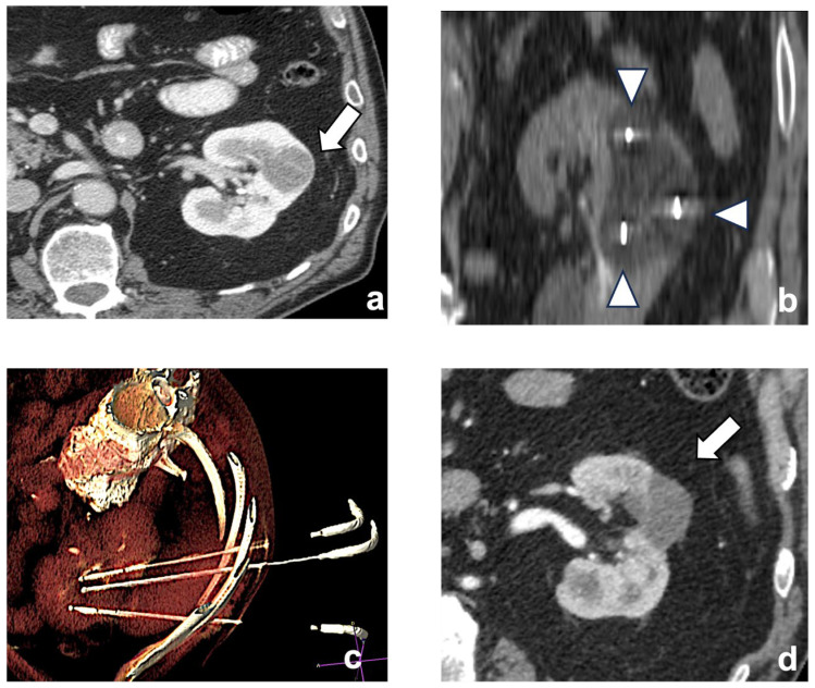
Figure 2.
A left chromophobe RCC (arrow), hypovascular compared to normal renal cortex (a). Three cryoablation probes (arrowheads) are placed in the tumor, creating an ice-ball ((b) coronal plane). A volume-rendering reconstruction represents the probes’ access and positioning in the renal mass (c). Post-contrast CT shows the positive outcome of the ablation (arrow (d)).

