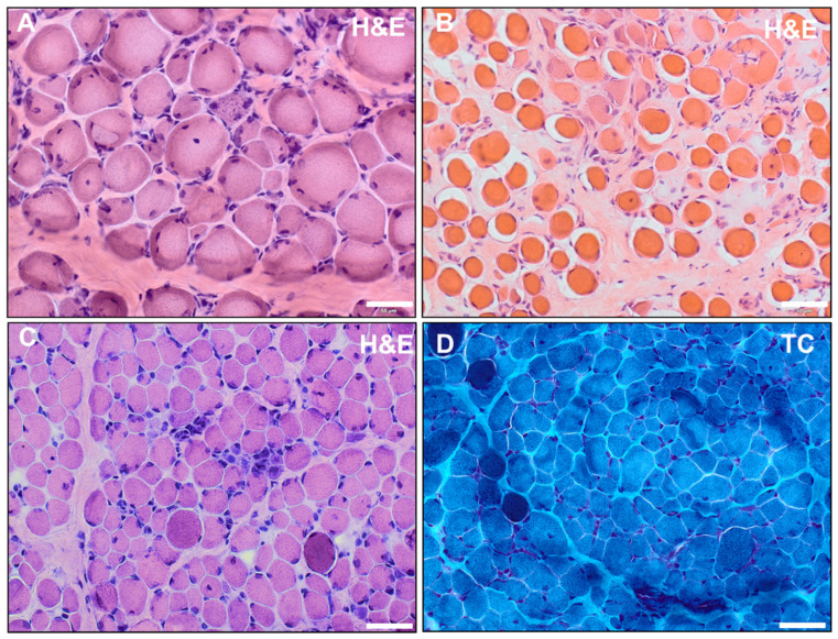
Figure 2.
(A,B) Hematoxylin and eosin (H&E) staining of quadriceps femoris muscle of patient 1 performed at the age of 2 (A) and of vastus lateralis muscle of patient 2 performed at 2.5 years (B) showed dystrophic alterations with enhanced presence of peri- and endomysial fat cells and connective tissue (indicative of fibrosis). Moreover, fiber size variations, hypertrophic and atrophic fibers, hypercontractive fibers, internal nuclei, as well as necrotic fibers were identified. (C,D) Histochemical staining of vastus lateralis muscle of patient 3 at the age of 1 revealed dystrophic alterations with endomysial fibrosis, fiber size variations, internal nuclei, hypercontractile fibers, and small infiltrates in H&E staining (C); in TC staining, the hypercontractive fibers appeared as darker-stained fibers (D) (scalebar: 50 µm).

