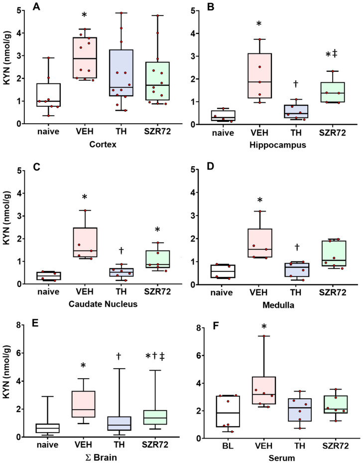
Figure 2.
Asphyxia-induced changes in brain KYN levels. Panel (A) shows cerebrocortical kynurenine (KYN) levels in the naive controls, as well as in the vehicle (VEH), hypothermia-treated (TH), and SZR72-treated (SZR72) groups subjected to asphyxia. Panels (B–E) show the values obtained from the hippocampus, the caudate nucleus, the medulla, and the combined data obtained from all assessed brain regions (ΣBrain), respectively. Panel (F) shows the combined serum data from the asphyxiated groups collected at 12–24 h after asphyxia, while baseline data (BL) were obtained from the VEH group collected before exposure to asphyxia. KYN levels increased significantly after asphyxia in all brain regions in the VEH group compared with controls. In contrast, KYN levels in the TH group were significantly reduced compared with the VEH group in the hippocampus, the caudate nucleus, and the medulla, while the trend was non-significant in the cerebral cortex. In fact, TH appeared to reduce KYN levels to control (naive) levels. SZR72 had a modest effect on asphyxia-induced KYN levels, but the trend was found significant only if all brain regions were considered together. Importantly, TH resulted in significantly lower KYN levels in the hippocampus and the caudate nucleus compared with SZR72 treatment. Lines, boxes, and whiskers represent the median, the interquartile range, and the 10th–90th percentiles, respectively. * p < 0.05, * significantly different from the naive, † from the VEH, ‡ from the TH group.

