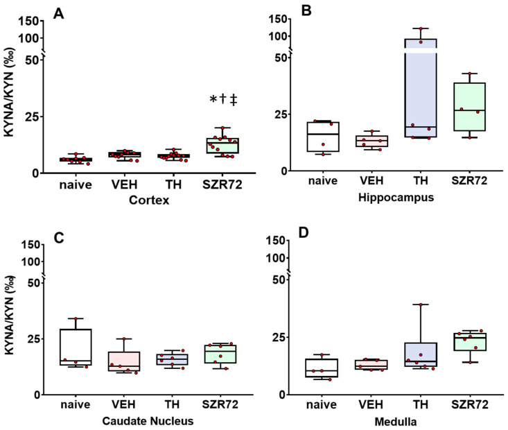
Figure 4.
Asphyxia-induced changes in brain KYNA/KYN ratios. Panel (A) shows cerebrocortical KYNA/KYN ratios in the naive controls, as well as in the vehicle (VEH), hypothermia-treated (TH), and SZR72-treated (SZR72) groups subjected to asphyxia. Panels (B–D) show the values obtained from the hippocampus, the caudate nucleus, and the medulla, respectively. KYN/KYNA ratio increased moderately, but significantly only in the cerebral cortex of the SZR72-treated group compared with all other groups. Otherwise, KYNA/KYN ratios were not significantly different in any other brain regions among the experimental groups. Lines, boxes, and whiskers represent the median, the interquartile range, and the 10th–90th percentiles, respectively. * p < 0.05, * significantly different from the naive, † from the VEH, ‡ from the TH group.

