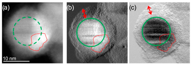
Figure 2.
(a) HAADF-STEM micrographs of the same single ION@UPy-NH2 indicating the crystalline NP core (green circle) and an overlapping smaller particle creating Moiré fringes (red outline); (b) DPCx (A–C) and (c) DPCy (B–D) images showing more clearly the NP core and the amorphous coating. (A–C) and (B–D) indicate different detector segments. The red arrow indicates the coating thickness.

