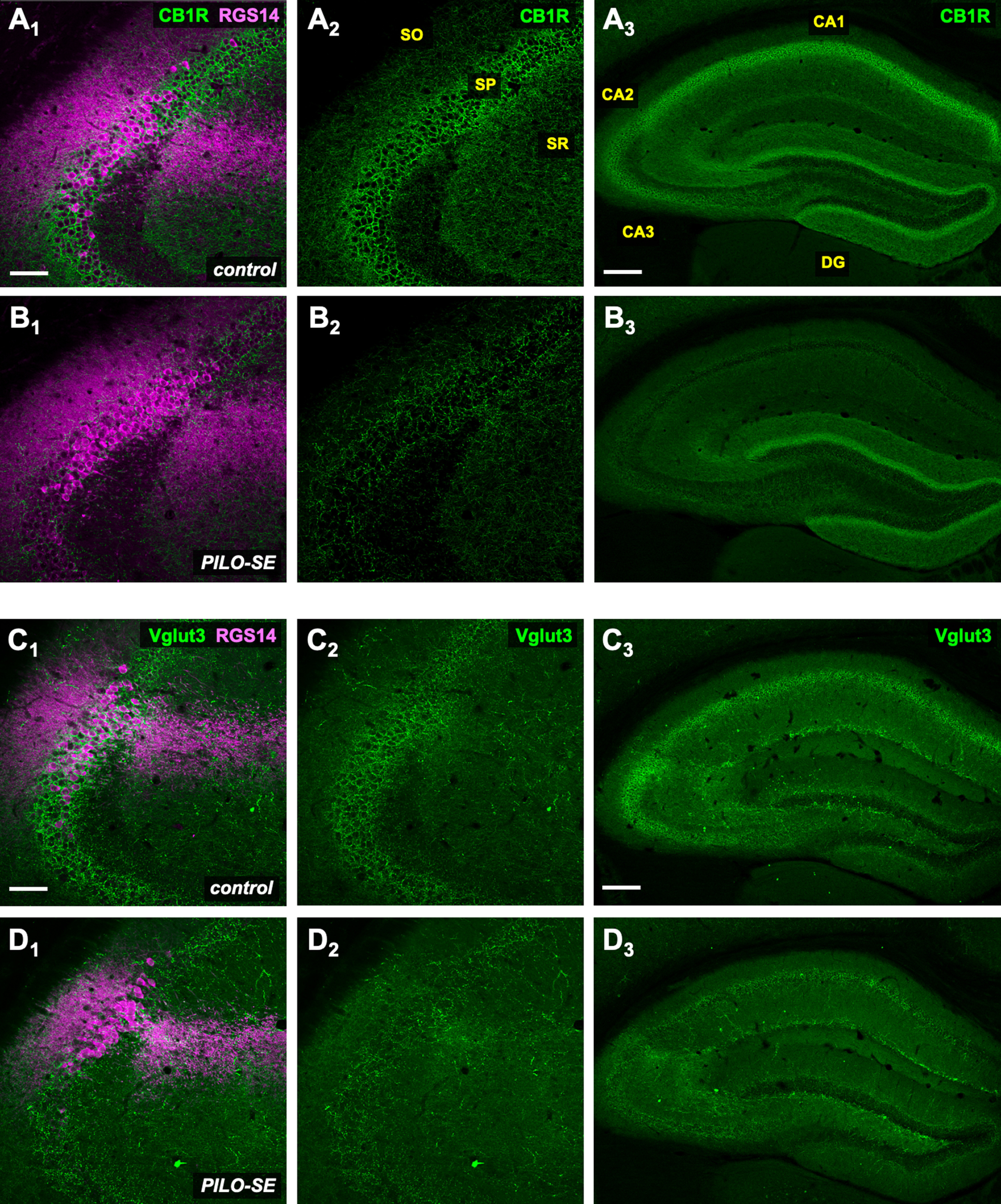
Figure 10.

Reduced labeling for molecular markers of CCK+ interneuron axon terminals in PILO-SE mice. A1–D3, Representative hippocampal sections from control (above) and PILO-SE (below) mice, stained for CB1R or VGluT3 (green) and RGS14 (magenta) to delineate the CA2 subfield, as indicated. Scale bars: left and middle panels, 100 µm; right panels, 400 µm.
