Figure 2.

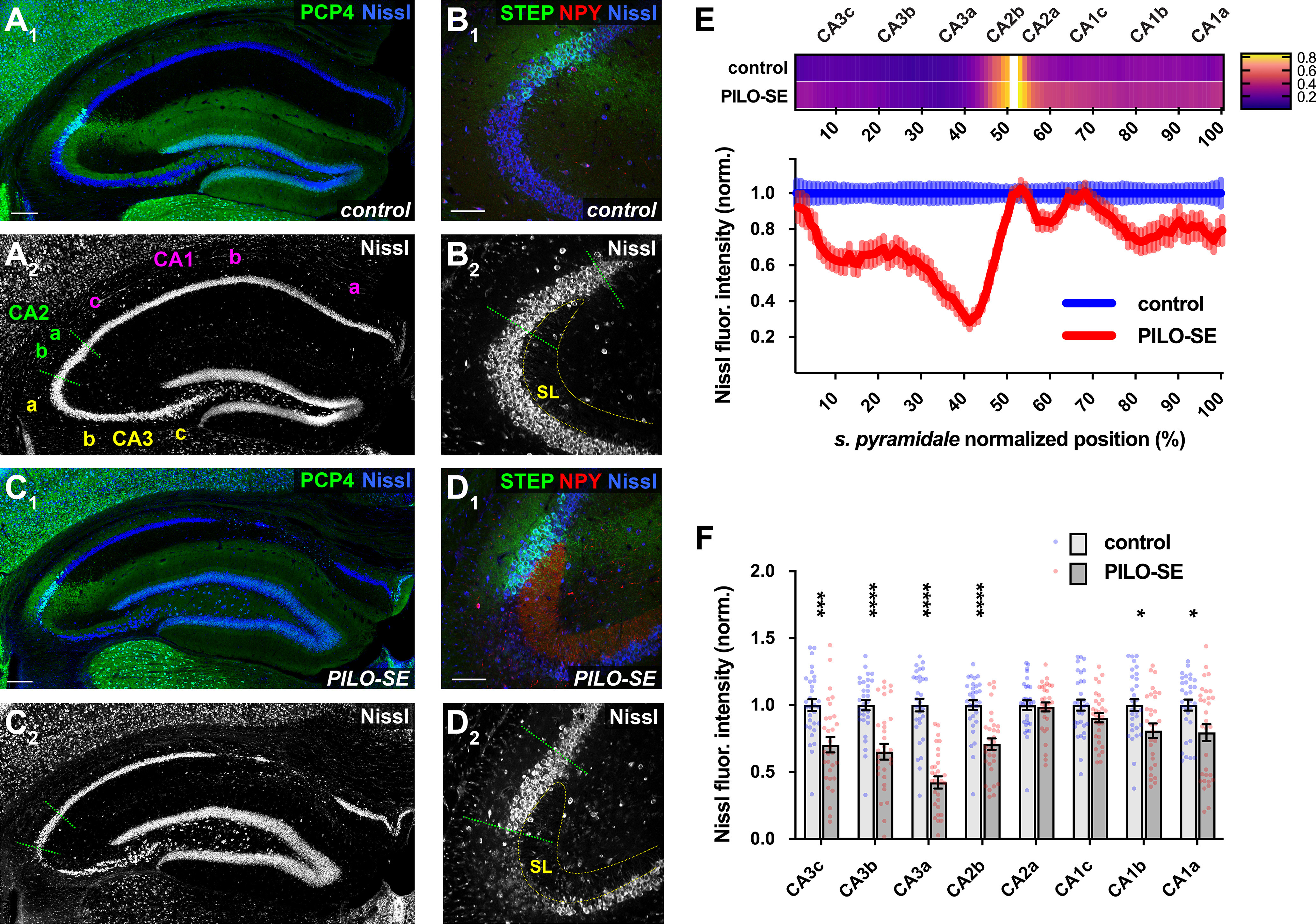
The CA2 subfield was resistant to mesial temporal sclerosis-like neurodegeneration. A1, A2, A representative section from a control mouse stained for PCP4 (green) to label CA2 PNs and for Nissl (blue and white). Scale bar, 200 µm. B1, B2, CA2 PNs (STEP, green) are located adjacent to CA3a at the distal end of the mossy fiber projection in SL. Neuronal somata are visualized with a stain for Nissl (blue and white); labeling for NPY is shown in red. Scale bar, 100 µm. C1, C2, Representative mesial temporal sclerosis-like damage in a section from a PILO-SE mouse stained for PCP4 (green) and Nissl (blue and white). Scale bar, 200 µm. D1, D2, NPY expression in the mossy fibers was visible in all sections from PILO-SE mice. Neuronal somata (Nissl, blue and white) are absent from CA3a and are largely preserved in CA2 (STEP, green). Scale bar, 100 µm. E, Above, a heatmap showing the normalized fluorescence intensity of CA2-specific markers (see Materials and Methods) along the proximal–distal axis of stratum pyramidale in sections from control and PILO-SE mice. Below, normalized Nissl fluorescence intensity across CA3, CA2, and CA1 was decreased significantly in CA3, along with the proximal region of CA2 (CA2b) and the distal portions of CA1 (CA1b, CA1a; n = 45 sections from 31 control mice, n = 56 sections from 31 PILO-SE mice). F, Measurement of the mean normalized Nissl fluorescence in each subregion confirms a characteristic pattern of neurodegeneration in PILO-SE mice with mesial temporal sclerosis-like damage, in which CA2a and CA1c are relatively resilient to neurodegeneration.
