Figure 3.

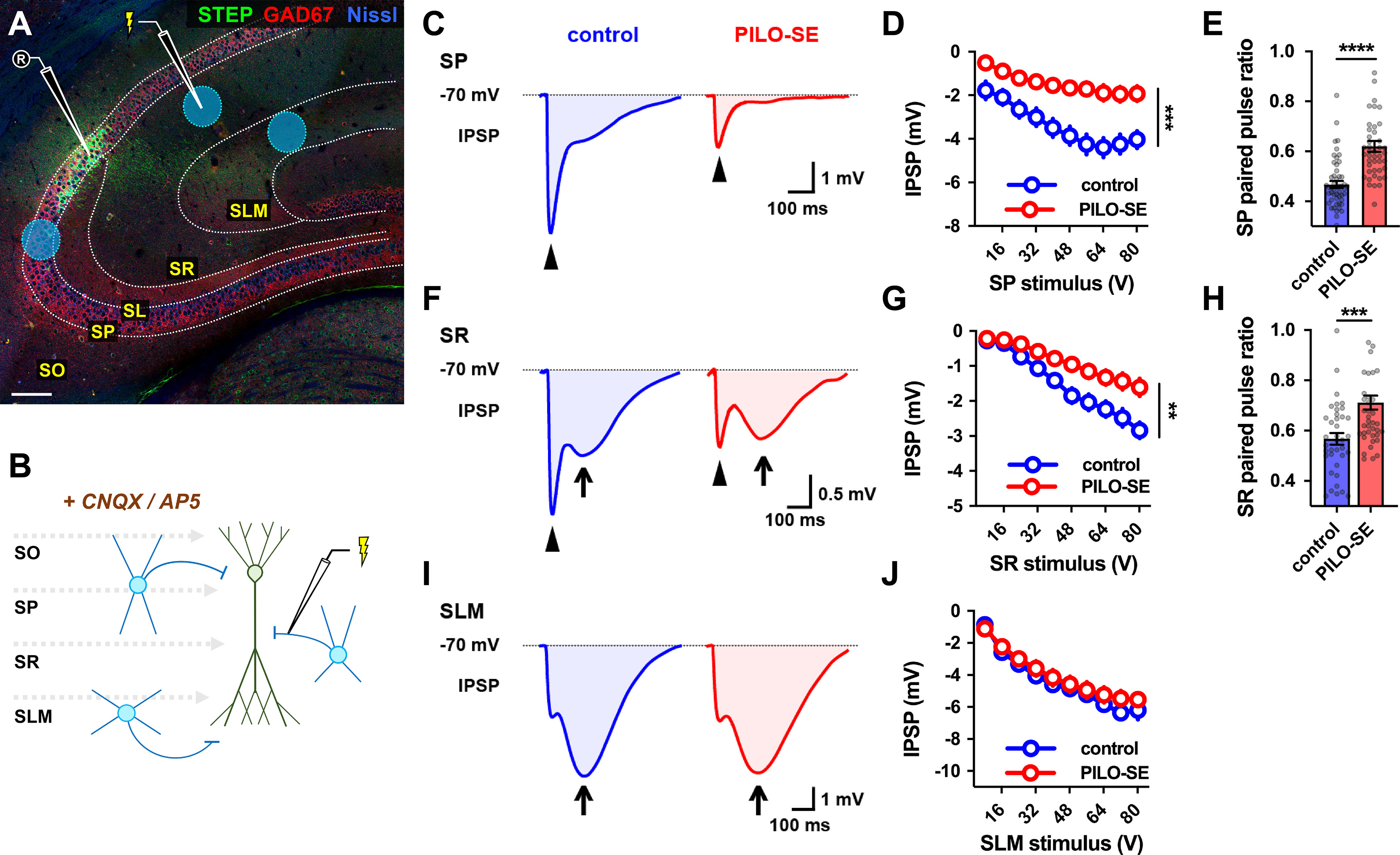
PILO-SE reduced CA2 PN perisomatic and proximal dendritic inhibition but did not alter inhibition at distal dendrites. A, A representative hippocampal section illustrating the stimulation locations for recruitment of monosynaptic inhibition in SP, SR, and SLM (blue circles). Stimulating electrode in SR is shown. CNQX and AP5 were added to block excitatory transmission and isolate IPSPs (see Materials and Methods). Scale bar, 125 µm. B, A circuit diagram illustrating the experimental configuration: a stimulation pipette located in SP, SR, or SLM evokes monosynaptic inhibition by directly activating local interneurons. C, Representative averaged IPSPs evoked in CA2 PNs by 64 V stimulation in SP in slices from control (blue) and PILO-SE (red) mice, with the fast peaks indicated with arrowheads. D, The peak hyperpolarization of the SP stimulation-evoked IPSP was significantly reduced in PILO-SE. E, The PPR of the IPSP evoked in SP was increased in PILO-SE mice. F, Representative averaged IPSPs evoked by 64 V stimulation in SR. The fast peaks are indicated with arrowheads, and the slow peaks with arrows. G, The peak fast hyperpolarization of the SR stimulation-evoked IPSP was significantly reduced in PILO-SE. H, The PPR of the SR stimulation-evoked IPSP was increased in CA2 PNs from PILO-SE mice. I, Representative averaged IPSPs evoked by 64 V stimulation in SLM, with the slow peaks indicated by arrows. J, The peak amplitude of the IPSP evoked by SLM stimulation, taking into account both fast and slow phases, was not significantly different between control and PILO-SE mice.
