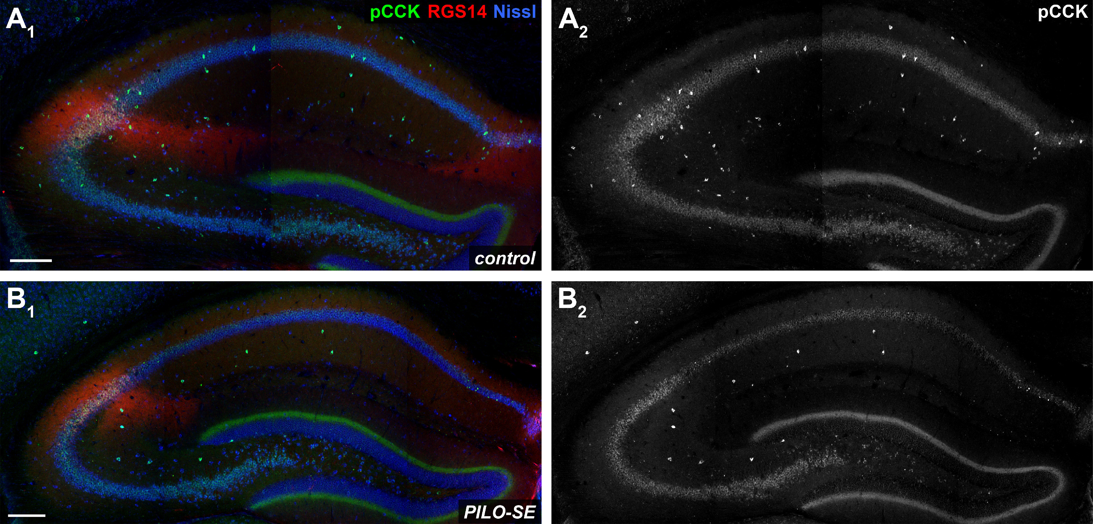
Figure 7.

PILO-SE was associated with a widespread decrease of pCCK+ interneurons. A1–B2, Representative hippocampal sections from control (A1, A2) and PILO-SE (B1, B2) mice, stained for pCCK (green and white) to visualize putative cholecystokinin-expressing interneurons, for Nissl to label neuronal somata (blue), and for RGS14 (red) to delineate the CA2 subfield. Scale bars, 200 µm.
