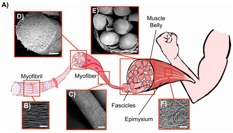
Figure 10.
Comparison between biological skeletal muscle and electrospun PU structures. (A) Schematic of the hierarchical structure of skeletal muscle. (B) Mat of aligned nanofibers. eA single nanofiber corresponds to a myofibril (scale bar = 20 µm). (C) Bundle of aligned fibers, corresponding to a myofiber (scale bar = 100 µm). (D) Cross-section of an aligned bundle, showing the parallel arrangement of the inner nanofibers (scale bar = 100 µm). (E) Cross-section of the hierarchical nanofibrous electrospun structure (HNES), compared to the cross-section of a biological muscle belly (scale bar = 150 µm). (F) HNES membrane, resembling the epimysium membrane that envelops the muscle belly (scale bar = 100 µm). Reprinted with permission from [115].

