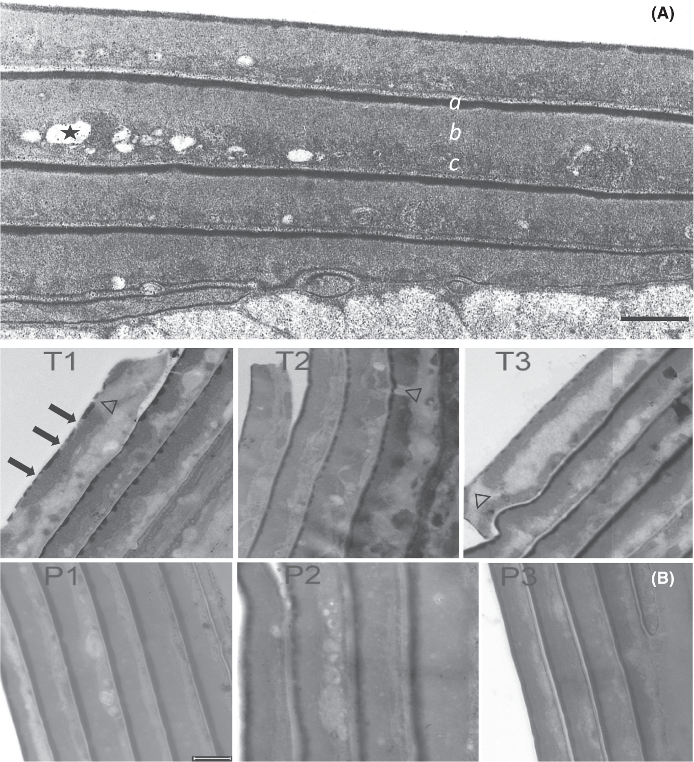
FIGURE 1.

Transmission electron micrographs. (A) Cuticle of normal hair incubated in 2% SDS – 25 mM DTT at pH 7.8 for 6 h. The shaft was fixed, stained, sectioned and examined by transmission electron microscopy as described.10 The dark layer on the outer edge of each cell (a) is called the marginal band or A-layer. The fine-grained layer immediately beneath the marginal band is called the exocuticle (b). The coarse-grained layer at the bottom edge of the cell is the endocuticle (c), in which remnants of intracellular organelles frequently appear embedded, some of which are extracted by the harsh detergent treatment (one indicated by *). (B) Hair shaft cuticle from TTD patients (T1, T2, T3), their respective parents (P1, P2, P3). Arrows in T1 point to discontinuities in the marginal band of the outermost cell; note the clear discontinuities in the next two inner cells. Discontinuities in the exocuticle layer in the outermost cell in T1 and T3 and an inner cell in T2 are denoted by Δ. Note the disorganized appearance of the endocuticle in cells in the TTD samples. Scale bars = 0.5 μm
