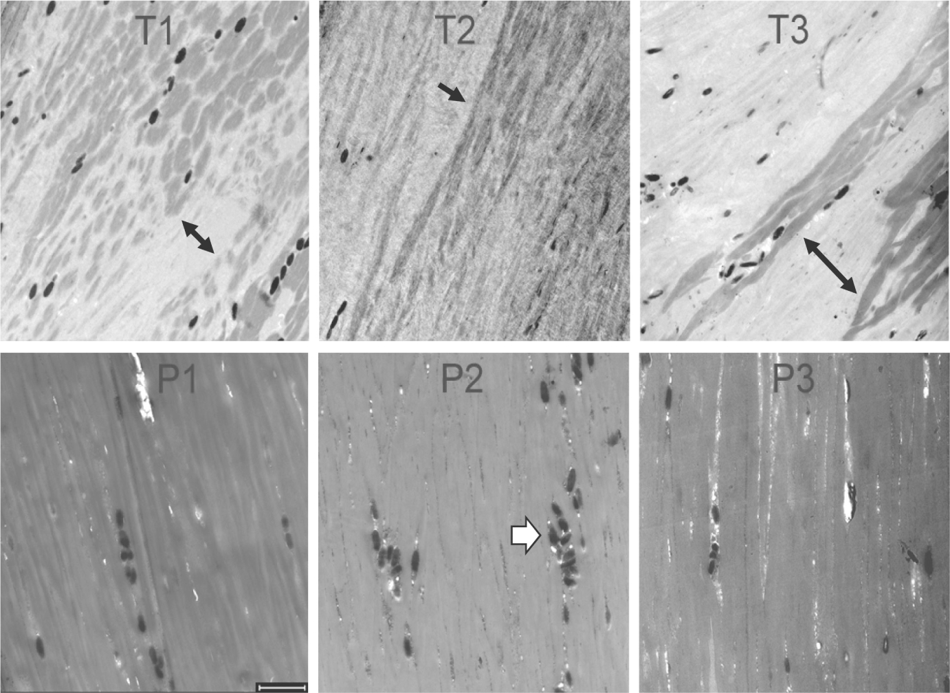
FIGURE 2.

Longitudinal sections of hair shafts (3 h incubation) from TTD patients (T1, T2, T3) and their parents (P1, P2, P3). Note the variegated staining patterns in T1, T2 and T3 (black arrows), which differ from the parental samples and from each other. The dark ellipsoids seen in all the samples (white arrowhead in P2) are melanosomes (0.4–0.9 μm diameter).28 Scale bar 2 μm
