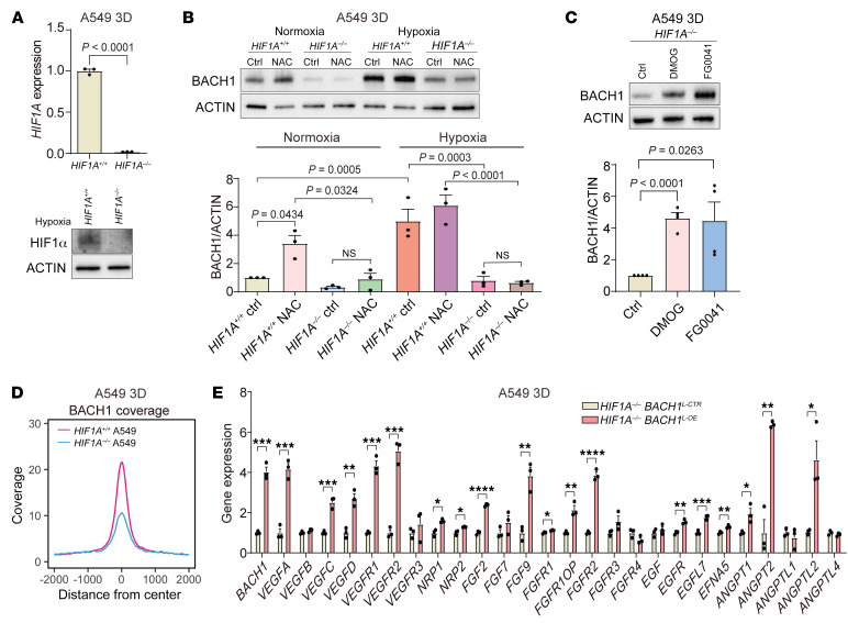
Figure 5. BACH1 increases angiogenesis gene expression in HIF1A-deficient lung cancer cells.
(A) HIF1A-knockout validation with RT-qPCR and Western blotting. (B) BACH1 protein levels by Western blotting in HIF1A–/– and HIF1A+/+ spheroids under normoxia and hypoxia and BACH1 levels by densitometry (n = 3 experiments). (C) BACH1 protein levels by Western blotting in HIF1A–/– spheroids incubated for 16 hours with prolyl hydroxylase inhibitors and BACH1 levels by densitometry (n = 4 experiments). (D) Genome-wide BACH1 CUT&Tag peak density plot of HIF1A+/+ and HIF1A–/– spheroids. (E) RT-qPCR of BACH1 and angiogenesis genes in HIF1A–/– spheroids with lentiviral BACH1 overexpression (BACH1L-OE) and controls (BACH1L-CTR) (n = 3 experiments). Data indicate the mean ± SEM. *P < 0.05, **P < 0.01, ***P < 0.005, and ****P < 0.001, by 2-tailed, unpaired Student’s t test (A and E) and 1-way ANOVA with Tukey’s post hoc test for multiple comparisons (B and C).

