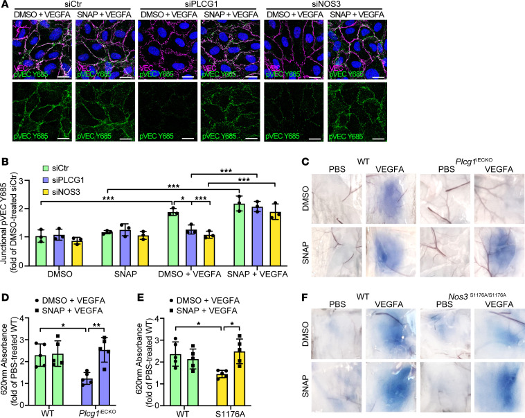
Figure 8. NO donor rescue of VE-cadherin phosphorylation and vascular leakage in the absence of PLCγ/eNOS signaling.
(A) Representative images of immunostainings with antibodies against VE-cadherin (VEC) and pVEC Y685 of HUVECs treated with VEGFA + DMSO or VEGFA + SNAP for 5 minutes, pretreated with siCtr, siPLCG1, or siNOS3. Scale bar: 20 μm. (B) MFI quantification of data from A shown as fold of DMSO-treated siCtr. n= 3 independent experiments, ≥3 fields of view/experiment. 1-way ANOVA. (C) Miles assay showing Evans blue leakage in the back skin after intradermal injection of PBS or VEGFA, combined with DMSO (control) or the NO-donor SNAP, in tamoxifen-treated Plcg1fl/fl; Cdh5-Cre– (WT), and Plcg1fl/fl; Cdh5-Cre+ (Plcg1iECKO) mice. (D) Quantification of extravasated Evans blue from C shown as fold change of PBS-treated WT mice, normalized to tissue weight; n = 5/genotype. 1-way ANOVA. (E and F) Quantification (E) and representative images (F) of Evans blue leakage in the back skin, in response to intradermal injection of PBS or VEGFA, cotreated with DMSO or SNAP, in Nos3+/+ (WT) and Nos3S1176A/S1167A (S1176A) mice; n = 5/genotype. 1-way ANOVA. Data represent mean ± SD. *P < 0.05, **P < 0.01, ***P < 0.001.

