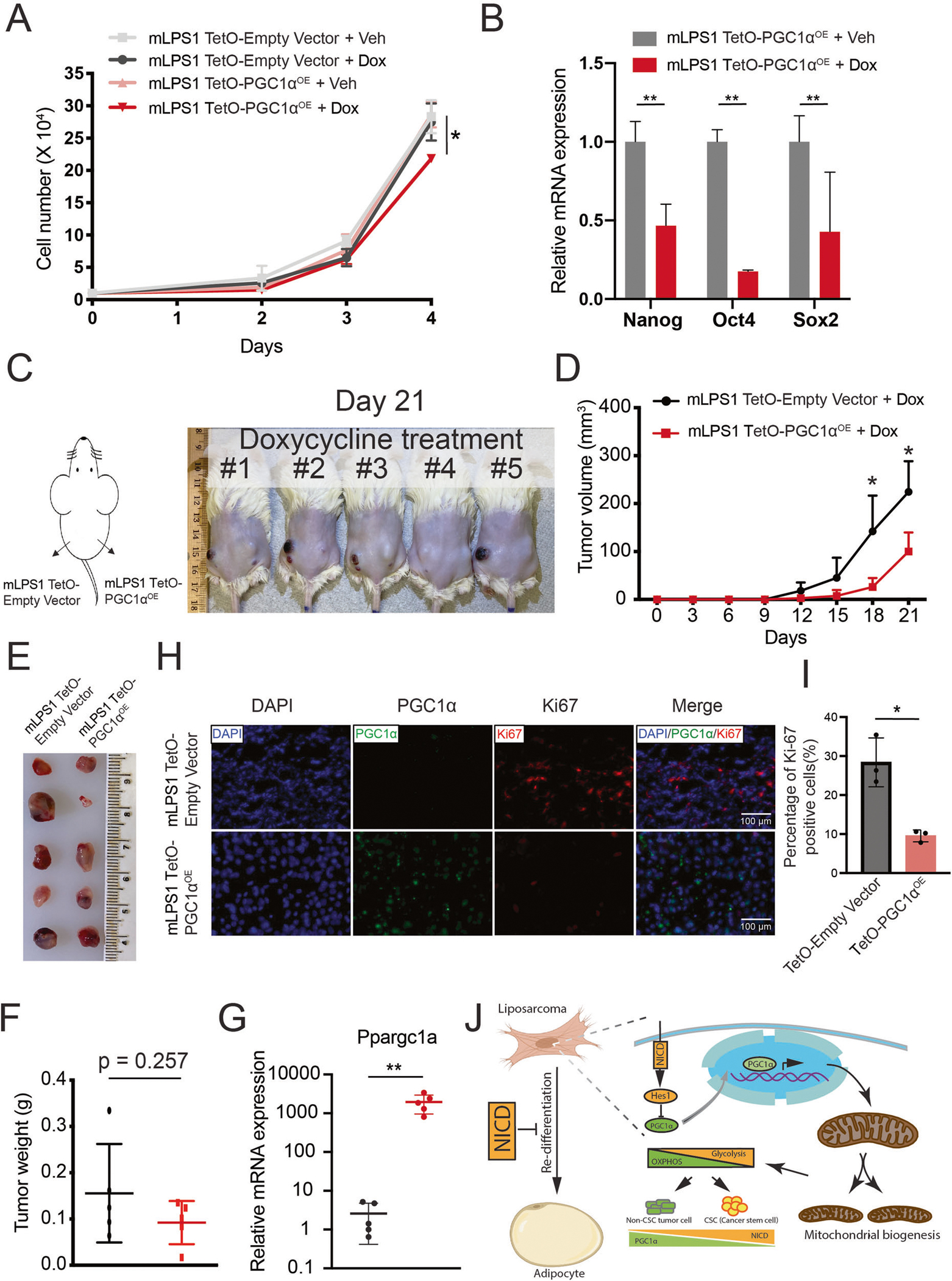
Fig. 8. PGC-1α expression attenuates mLPS1 cell proliferation and tumorigenesis.

A Cell proliferation of TetO-Empty vector and TetO-mPGC1αOE stable transfected mLPS1 cells treated with doxycycline (dox) or vehicle control (veh) (n = 3). B RT-qPCR analysis of the relative mRNA levels of cancer stem cell markers in Veh or Dox treated TetO-mPGC1αOE mLPS1 cells (n = 3). C The mLPS1 TetO-mPGC1αOE cells and mLPS1 TetO-Empty vector cells were subcutaneously transplanted into NRG mice and treated with Dox via drinking water (n = 5) for 21 days. D Tumor growth curves based on tumor volume calculation obtained from caliper measurement. E Morphology and (F) average weight of the transplanted tumors. G RT-qPCR analysis of PGC-1α expression in the transplanted tumors. H Immunofluorescent staining shows the relative expression of PGC-1α and Ki67 in the mLPS1 TetO-Empty vector and the mLPS1 TetO-mPGC1αOE allograft tumors. Nuclei were stained by DAPI. I Quantitation of Ki67 positive cells in H. Data are represented as mean ± SD. *P < 0.05, **P < 0.001. J Graphic illustration of how Notch signaling regulates cancer cell differentiation and mitochondrial function in liposarcoma.
