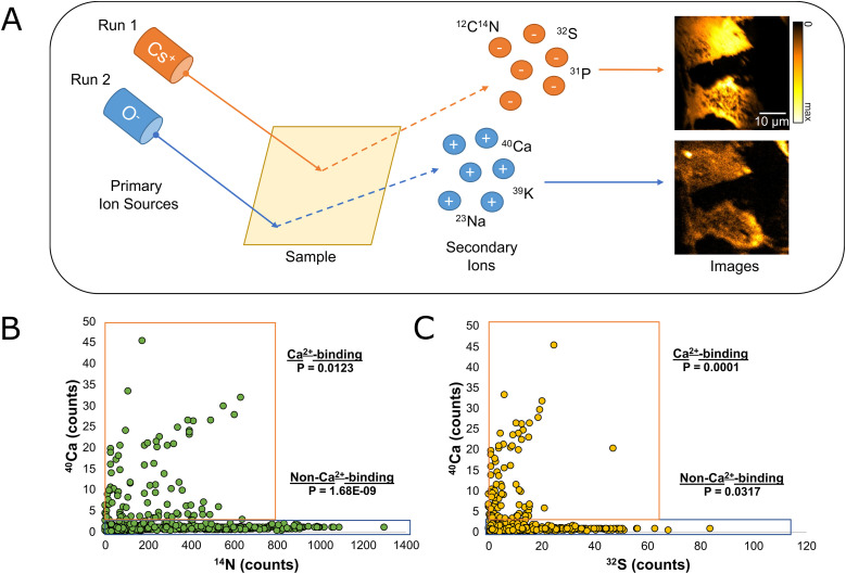
Figure 1. Comparison between calcium, sulfur, and nitrogen peaks as observed by nanoscale secondary ion mass spectrometry.
(A) Nanoscale secondary ion mass spectrometry is a chemical-imaging technique which uses an ion source (Cs+ or O−) to produce a charged primary ion beam. When the beam strikes the surface of the sample, secondary ions of the opposite charge are created and measured. To analyze both 14N and 40Ca in our samples, it was necessary to first analyze the samples using the Cs+ source. Once all samples were analyzed using Cs+, the instrument was switched over to a radio-frequency RF source, and 40Ca was measured in a second run. (B) The intensity of the 40Ca peak at various points from each image is compared with the intensity of the 14N peak at the same location. The data are divided into two groups: a Ca2+-binding group (40Ca > 2, N = 104, P = 0.0123), and a non-Ca2+-binding group (40Ca < 2, N = 462, P = 1.68 × 10⁻⁹). (C) The intensity of the 40Ca peak at various points from each image is compared with the intensity of the 32S peak at the same location. The data are divided into two groups: a Ca2+-binding group (40Ca > 2, N = 104, P = 0.0001), and a non-Ca2+-binding group (40Ca < 2, N = 462, P = 0.0317).
Source data are available for this figure.

