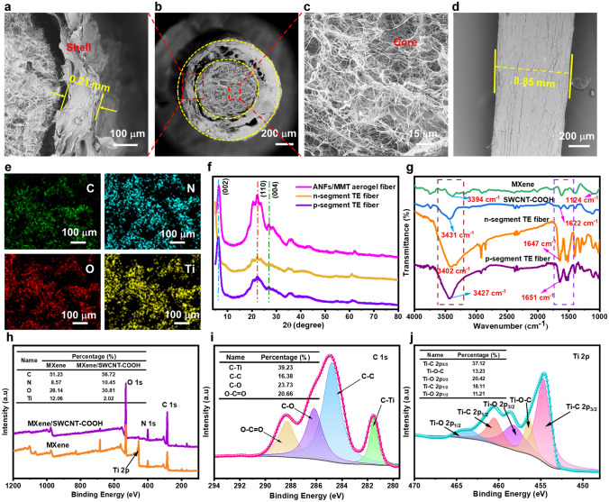
Fig. 2.
Microstructural characterizations of p–n segment core–shell TE fibers. a Cross-sectional SEM image of TE fiber shell. b Cross-sectional SEM image of the p–n segment TE fibers. c SEM image of p-segment TE fiber core material. d Surface morphology of p–n segment TE fiber. e EDX mapping images of p-type core material in TE fiber. f XRD patterns of pure ANFs/MMT fiber, p-segment and n-segment TE fibers. g FTIR spectra of MXene, SWCNT-COOH, p-segment TE and n-segment TE fibers. h Wide-scan XPS spectra of n-type MXene and p-type MXene/SWCNT-COOH samples. High-resolution XPS spectra of i C 1s and j Ti 2p for MXene/SWCNT-COOH sample

