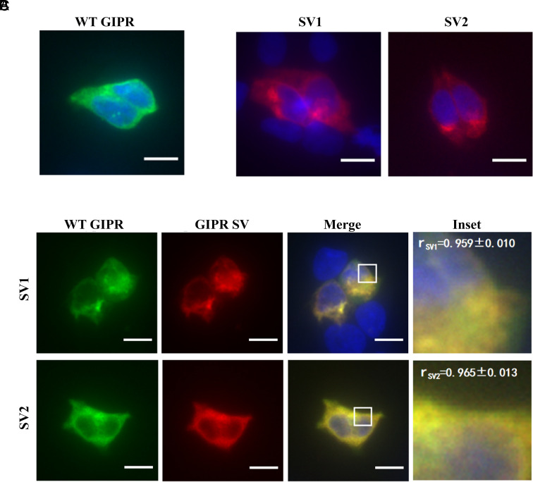
Fig. 6.
Colocalization of GIPR and its SVs. Immunofluorescence staining of HEK293T cells transfected with GIPR-HA (A) or each SV-FLAG (B) alone. To estimate their colocalization, cotransfection of GIPR and individual SVs (C) was performed at a ratio of 1:3 (green, GIPR-HA; red, SV-FLAG; yellow, merge). Data show representative results from three independent experiments. The inset demonstrates the overlapping positions of GIPR and SVs in the cells. Cells were observed by DeltaVision™ Ultra. (Scale bar, 10 μm.) Pearson’s correlation coefficients (r) were determined using the Image J colocalization threshold plugin. Values are means ± SD.

