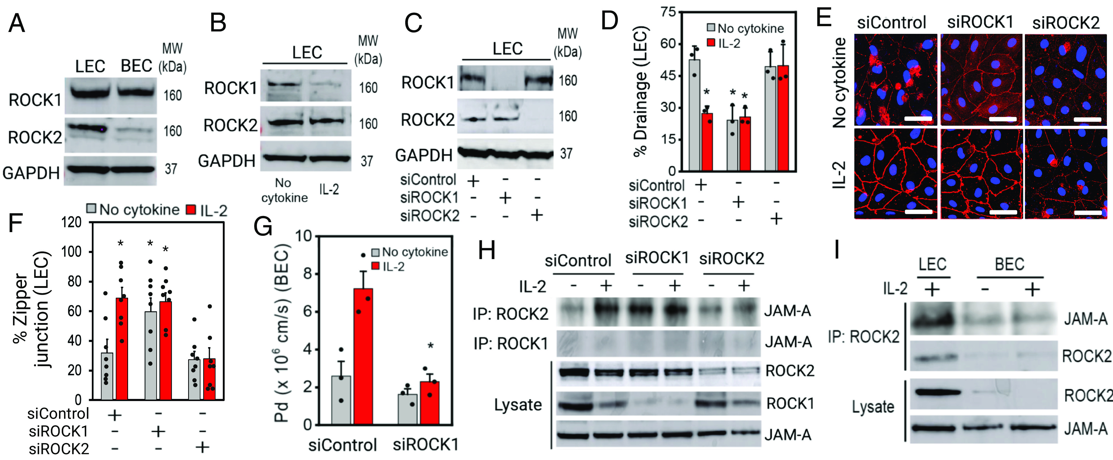
Fig. 3.

ROCK2 forms a unique tight junction complex in inflamed LECs, but not in BECs. (A) Western blot showing basal levels of ROCK1/2 expression in LECs and BECs. (B) Western blot showing ROCK1 downregulation in inflamed LECs. (C) siRNA-mediated knock-down of ROCK1 and ROCK2 in LECs. (D) Percent lymphatic drainage by LEC knocked-down ROCK1 or ROCK2 in normal or IL-2 condition (BODIPY-C16 fatty acid drainage) (N = 3). (E) VE-cadherin images of the engineered lymphatics with or without ROCK1/2 knock-down in normal or IL-2 condition. (F) Percent zipper junction in engineered lymphatics with or without ROCK1/2 knock-down in normal or IL-2 condition (n = 8, N = 3). (G) Permeability coefficient (Pd) of the engineered blood vessels with or without ROCK1 knock-down in normal or IL-2 condition (N = 3). (H) Immunoprecipitation data showing ROCK2 interactions with junctional adhesion molecule-A (JAM-A) in inflamed LECs or in ROCK1 kd LECs. (I) Immunoprecipitation data showing that ROCK2-down-regulating BEC does not form ROCK2/JAM-A protein complex in normal or IL-2 condition. [Scale bars (E), 50 μm.] *P < 0.05 indicates statistical significance.
