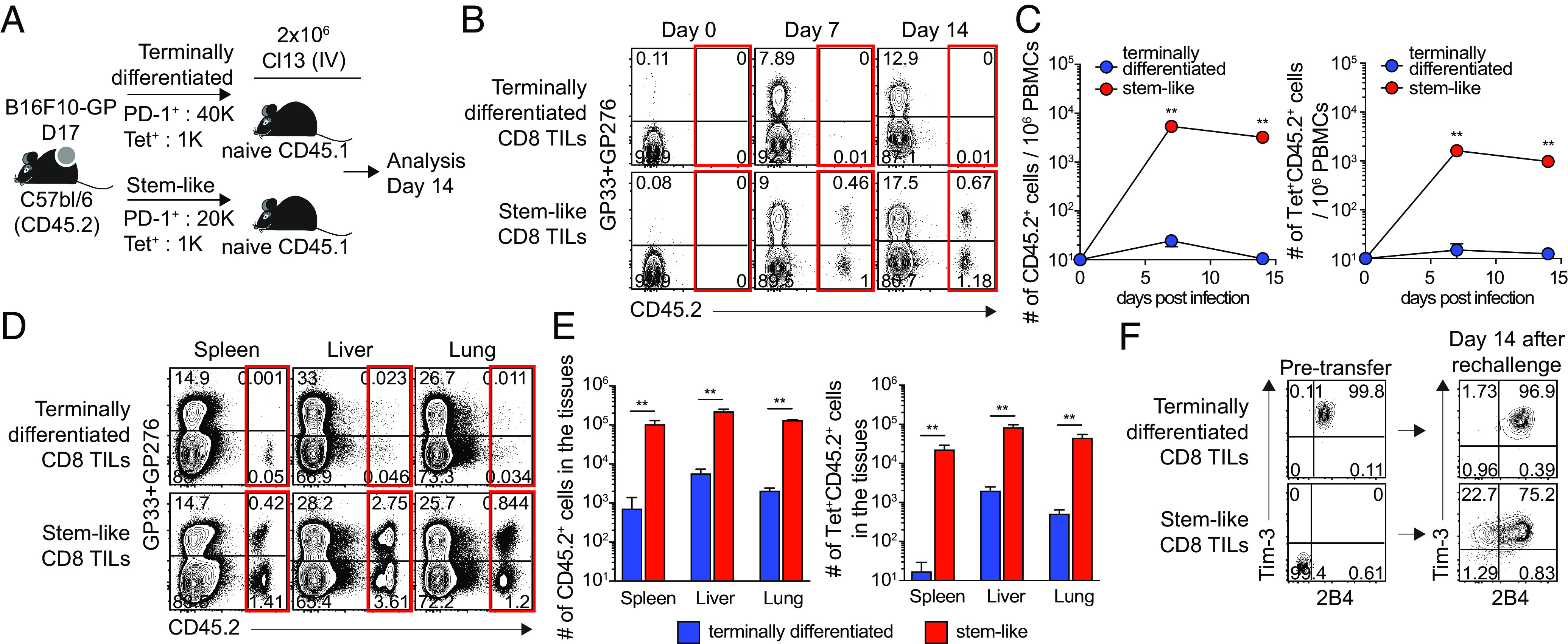
Fig. 2.

Proliferative potential of stem-like CD8 T cells isolated from tumor-bearing mice upon antigen reexposure. (A) Experimental setup illustrating transfer of congenically marked Tim-3+2B4+ PD-1+ (terminally differentiated) and Tim-3−2B4− PD-1+ (stem-like) CD8 T cells isolated from B16F10-GP-bearing mice (day 17) into naive Ly5.1 recipient mice, followed by LCMV Cl-13 challenge. (B and C) Representative FACS plots (B) and kinetics of total donor cells and donor GP33/GP276-specific CD8 T cells (C) in the blood (D and E) Representative FACS plots (D) and the number of total donor cells and donor GP33/GP276-specific CD8 T cells (E) in the tissues at day 14 post the challenge. (F) Phenotypic analysis of donor CD8 TIL subsets before and after the transfer followed by Cl-13 challenge. Data were representative of two independent experiments (n = 4/experiment). Graph shows the mean and SEM. Student’s t test, where **P < 0.01.
