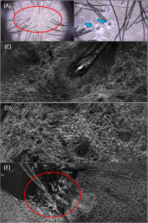
FIGURE 2.

Trichoscopic image of patient with classic type of lichen planopilaris. Perifollicular scaling (20x magnification, red oval and 70x magnification blue arrows) is visible on trichoscopic picture (A,B). LC‐OCT image in a horizontal mode showed scarring around the hair follicle (C). LC‐OCT image in a horizontal mode revealed increased number of white, ill‐defined, coarse dermal fibres corresponding to the scarring (D). LC‐OCT image in a horizontal mode showed perifollicular hyperkeratosis (F).
