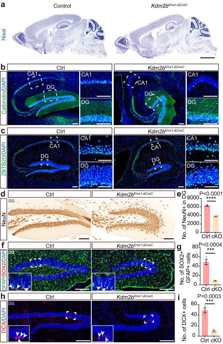
Fig. 1. Deletion of the KDM2B-CxxC causes hippocampal hypoplasia.
a Representative images showing Nissl staining on sagittal sections of adult control and Kdm2bEmx1-ΔCxxC brains. b, c Immunofluorescent (IF) staining of Calbindin (b) and ZBTB20 (c) on sagittal sections of adult control (left) and Kdm2bEmx1-ΔCxxC (right) hippocampi. Nuclei were labeled with DAPI (blue). Boxed CA1 and dentate gyri (DG) were enlarged on the right. d Immunohistochemical (IHC) staining of NeuN on sagittal sections of adult control (left) and Kdm2bEmx1-ΔCxxC (right) DG. f, h Double immunofluorescence of GFAP (green) and SOX2 (red) (f) and single immunofluorescence of DCX (h) on sagittal sections of adult control (left) and Kdm2bEmx1-ΔCxxC (right) DG. Nuclei were labeled with DAPI (blue). Boxed regions were enlarged on bottom-left corners. Arrows denote GFAP + SOX2+ or DCX+ signals in the subgranule zone (SGZ). e, g, i Quantification of NeuN+ cells in the DG (e), GFAP + SOX2+ cells in the SGZ (g) and DCX+ cells in the SGZ (i). n = 3 for control brains and n = 4 for Kdm2bEmx1-ΔCxxC brains. Similar results were obtained for 3 control brains and 4 Kdm2bEmx1-ΔCxxC brains (a–c). Data are represented as means ± SEM. Statistical significance was determined using an unpaired two-tailed Student’s t-test (e, g, i). ***P < 0.001; ****P < 0.0001. Scale bars, 2 mm (a), 100 μm (b–d), 200 μm (f, h), 50 μm (c CA1 and DG).

