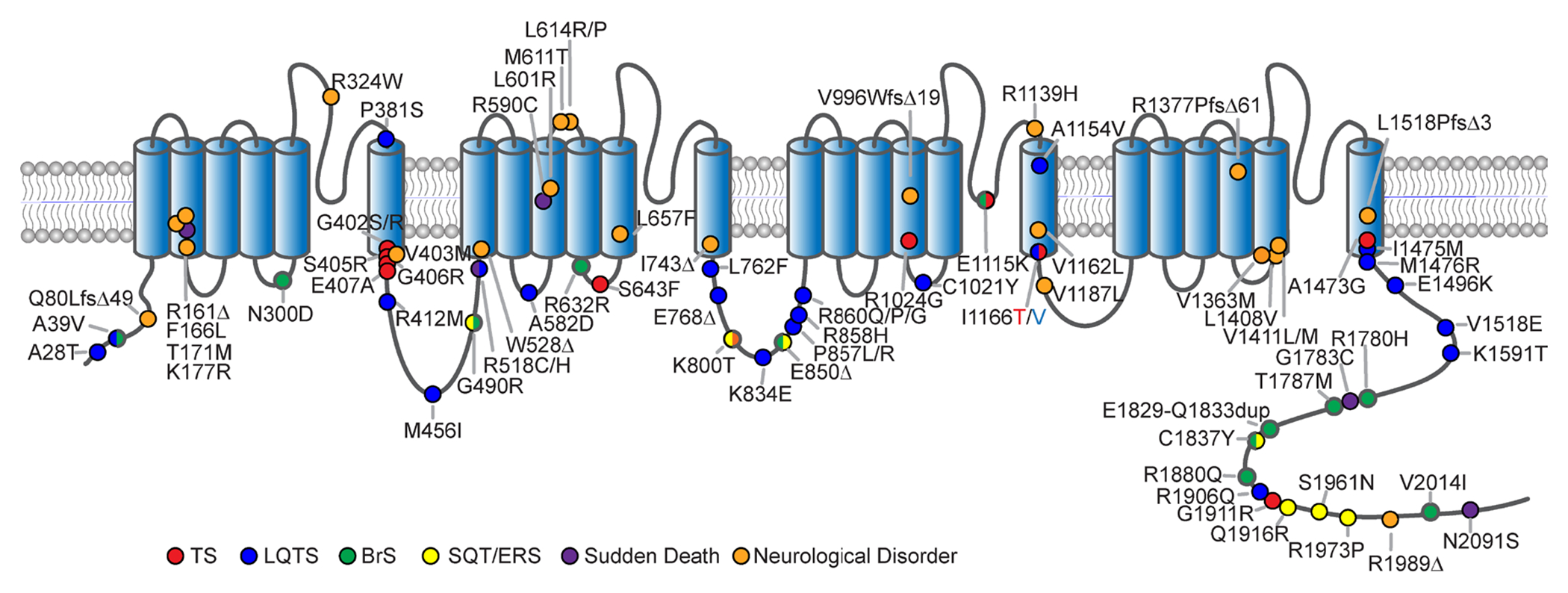
Figure 1. CACNA1C mutations.

Cartoon depicting the membrane topology of the pore forming subunit of CaV1.2 indicating the locus of CACNA1C mutations across the channel. Phenotypes associated with each mutation are colored accordingly.

Cartoon depicting the membrane topology of the pore forming subunit of CaV1.2 indicating the locus of CACNA1C mutations across the channel. Phenotypes associated with each mutation are colored accordingly.