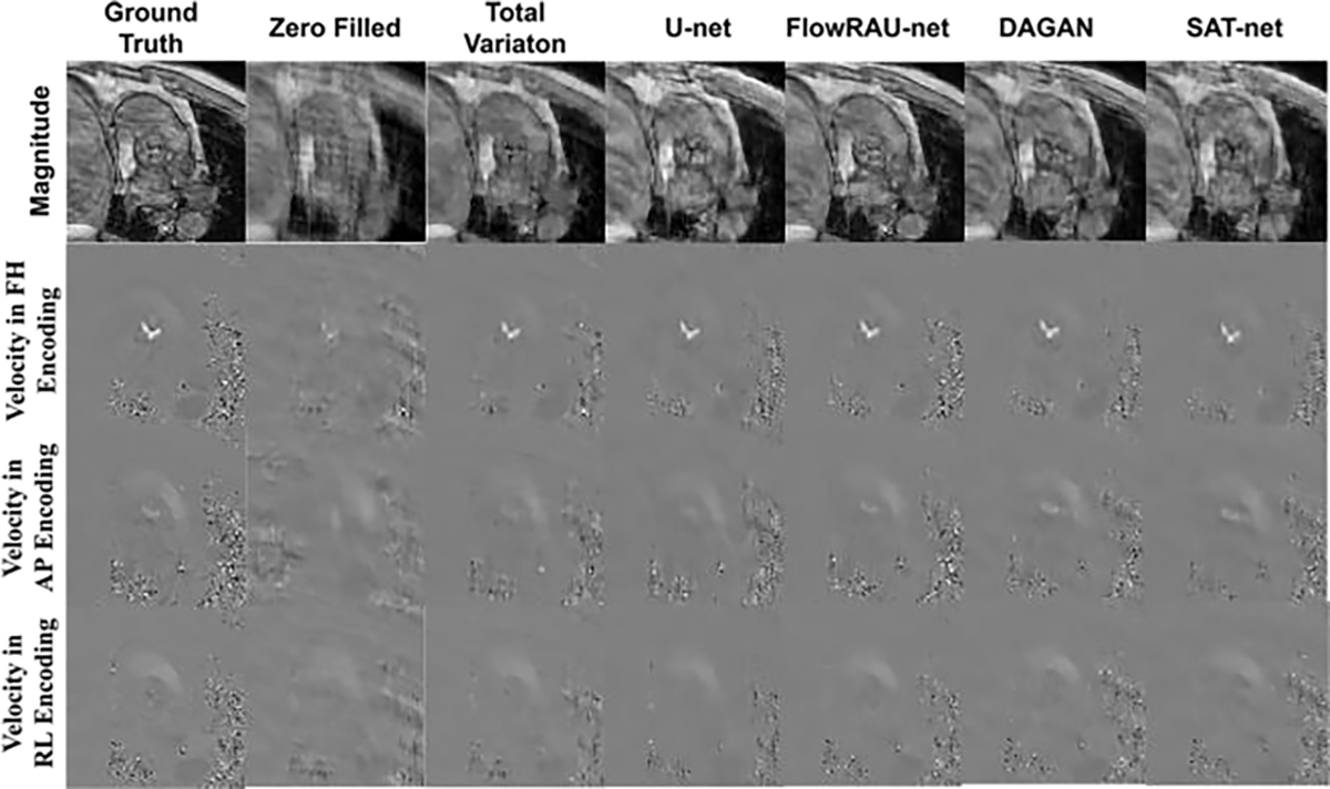
Fig. 6:

Comparison of 4D Flow reconstructed images for various techniques in one patient in-vivo. The first row shows magnitude image (in one encoding direction). The second, third, and fourth row shows velocity mapped image at FH, AP and RL direction from the reference image, the zero- filled reconstructed image, and the image reconstructed by U-net, TV regularization, DAGAN, SAT-net and proposed FlowRAU-net method. The images are in the peak systole phase of the cardiac cycle and is exactly at the location of the aortic valve.
