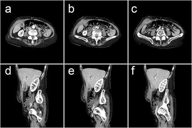
Figure 1.

Representative CT images showing (a–c) axial and (d–f) sagittal views of a 47 × 23 × 66 mm complex collection within the right anterior abdominal wall, consisting of fluid and phlegmonous components. This collection is contiguous with a thick-walled gallbladder that contains calculi. These findings are consistent with an abdominal wall abscess complicating acute-on-chronic cholecystitis with extraperitoneal perforation.
