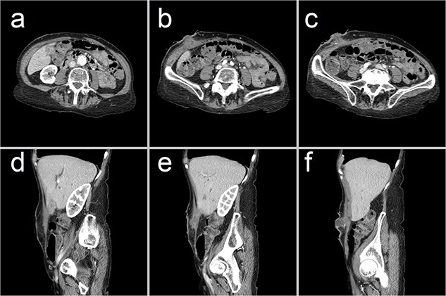
Figure 3.

Representative CT images showing (a–c) axial and (d–f) sagittal views of the CCF, forming a communicating tract that extends from the gallbladder fundus, through the anterior abdominal wall and onto the skin. There has been interval collapse of the gallbladder, whose wall remains thickened with mucosal hyperenhancement, in keeping with chronic cholecystitis. Hyperenhancing gallstones are visible along this fistula tract.
