
Figure 1.
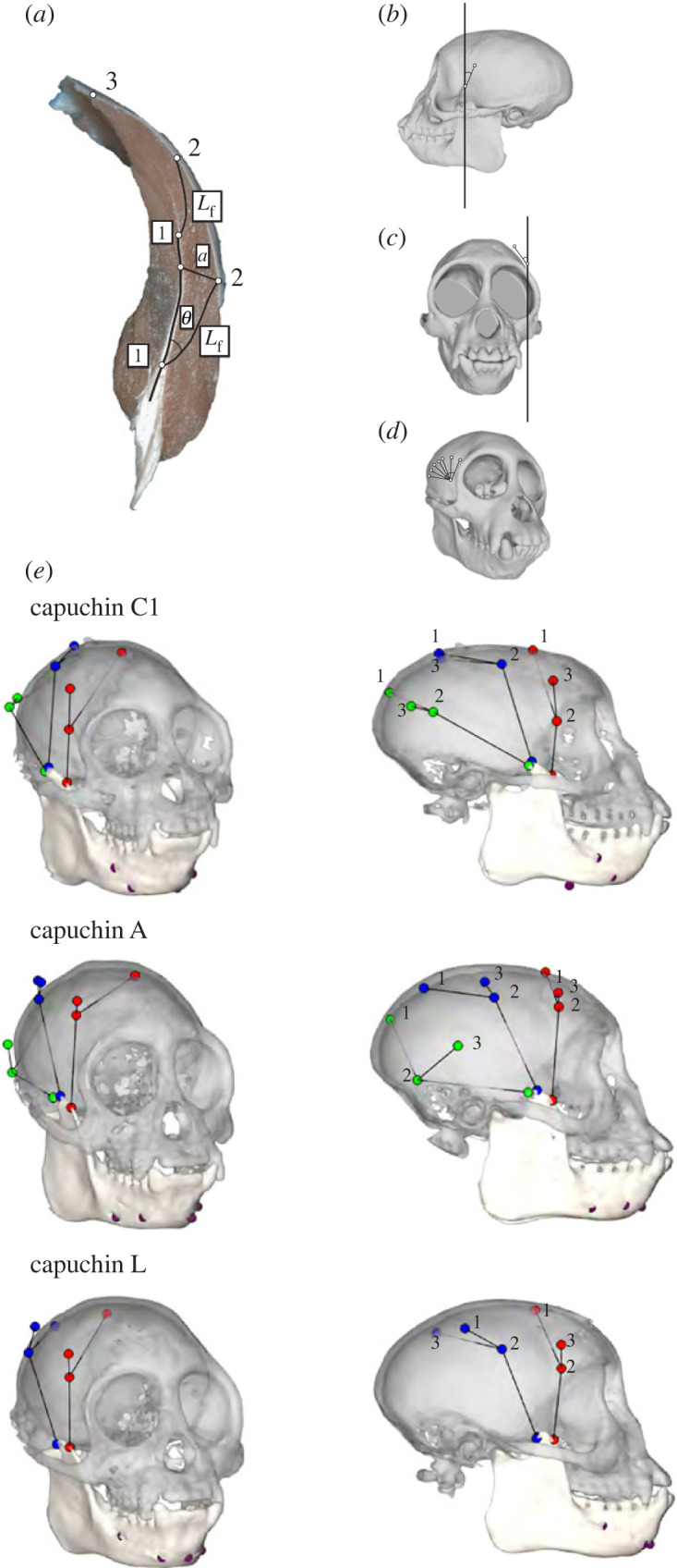
(a) A coronal section of a capuchin right temporalis in anterior view. Static muscle fibre length (Lf) and pinnation angle (θ) were measured between the central myotendinous junction and the fibre superficial termination. For dynamic muscle architecture, tantalum beads were sutured to the central myotendinous junction of a single fascicle (1), the superficial termination of that fascicle (2), and at the temporal fascia line directly superior to beads 1 and 2 (3). Fascicle angles were measured by passing planes between makers 1 and 3 in a coronal plane for (b) sagittal fascicle angle, and in the sagittal plane for (c) coronal fascicle angle. (d) Three-dimensional angle was measured as the change in angulation between markers 1–3 during a gape cycle. (e) Locations of the fascicle markers in the superficial anterior temporalis (SAT, in red), superficial middle temporalis (SMT, in blue) and superficial posterior temporalis (SPT, in green) for capuchin A, C1 and L.