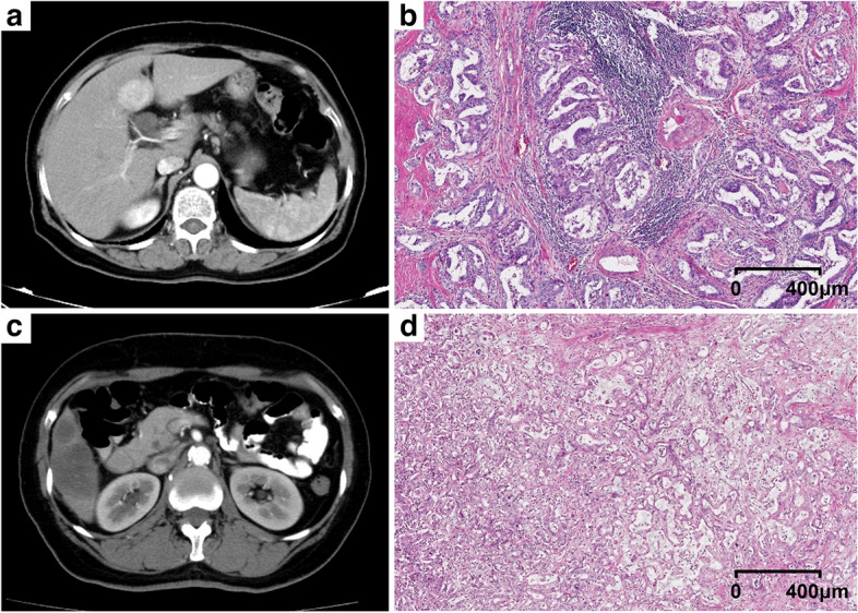
Fig. 5.

Two representative cases with TLSs-positive (a, b) and TLSs-negative (c, d) tumors were presented. For the TLSs-positive tumor, the arterial phase of enhanced CT showed diffuse hyperenhancement (a); the rad-score was 0.43; the nomogram score was 2.83; the pathological hematein-eosin saffron-stained slide (b) showed the immune infiltration in TLSs; the RFS of the patient was 25.30 months. For the TLSs-negative tumor, the arterial phase of enhanced CT showed diffuse hypoenhancement (c); the rad-score was −0.78; the nomogram score was −1.63; the pathological hematein-eosin saffron-stained slide (d) showed diffuse tumor cells and no TLSs in the tumor; the RFS of the patient was 1.20 months
