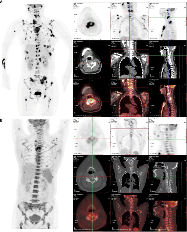
Figure 1.
(A) The 18F-FDG PET/CT image on January 20, 2022: Right frontal bone, jaw bone, occipital slope, right humerus, sternum, bilateral clavicles, bilateral shoulder blades, bilateralmultiple ribs, extensive cervical/thoracolumbar vertebrae appendages, pelvic bones, and bilateralfemurs were involved. Some lesions were associated with soft tissue masses, with significantlyincreased radiotracer uptake. The size of the soft tissue masses in the sternum region wasapproximately 5.5 cm×2.9 cm×3.8 cm, and the SUVmax was 16.8. Increased and enlargedlymph nodes were seen in the right parotid gland region, right supraclavicular region, and leftarmpit, with increased radiation uptake. The largest lymph node was in the left armpit(approximately 1.6 cm × 1.4 cm in size, SUVmax 15.6.). (B) The 18F-FDG PET/CT image of thepatient after receiving two courses of chemotherapy on March 19, 2022. Compared with the (A) image, in the (B) image shown, there was a significant reduction or disappearance of radioactivityuptake in multiple bones and multiple lymph nodes, including the virtual disappearance of thesoft tissue mass in the sternal region, and a reduction of radioactivity uptake in the adjacentsternum compared with the previous one (SUVmax: 10.6). The left axillary lymph node lesiondisappeared without significant radioactivity uptake. Furthermore, bone fractures exist, and thebone marrow metabolic activity is increased (likely due to the effect of colony-stimulatingfactors).

