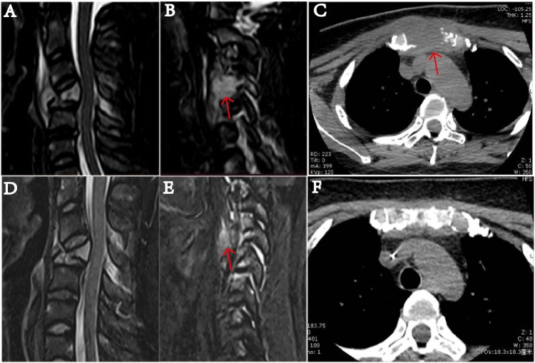
Figure 2.
MRI examination of the cervical vertebra of the patient on January 20, 2022. (A) shows slope, C4, T1 vertebrae, and ancillary multiple bony lesions with pathologic fracture of the C4 vertebrae. (B) shows a 1.49 mm ×1.54 mm mass (indicated by the arrow) near the C4 cone, and the C4 vertebral body is compressed and flattened and shows pathological fracture. (C) The sternum is fractured, and there is a 37.41 mm × 31.14 mm soft tissue mass (indicated by the arrow). On March 19, after receiving two courses of chemotherapy, the patient showed destruction of multiple bones and a cervical spine fracture on MRI (D–F). In part (E), the C4 paravertebral mass is significantly reduced (indicated by the arrow). In part (F), the soft tissue mass causing the sternum bone fracture is essentially undetectable.

