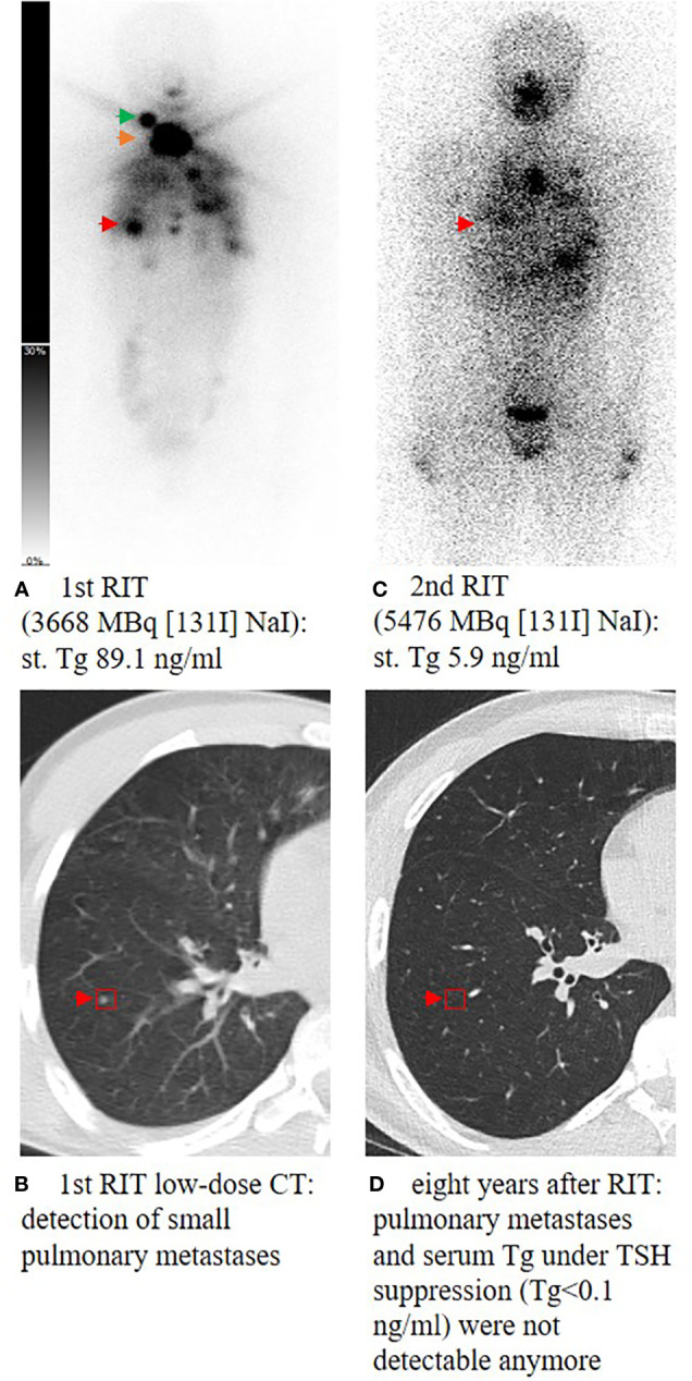
Figure 1.

Planar whole-body scintigraphy and CT scan after the first radioiodine therapy (RIT) and during follow-up. A 14-year-old male patient (A) presenting with high residual thyroid bed uptake (orange arrow), an iodine-positive cervical lymph node metastasis in level II on the right side (green arrow), and multiple iodine-positive pulmonary metastases (red arrow) (B). Post-therapeutic scan of the second RIT showed ablation of the remnant thyroid tissue and significant uptake of pulmonary metastases (C). During follow-up, TSH was suppressed consequently; 1.6 years after the last RIT, Tg decreased to <0.1 ng/mL and pulmonary metastases could not be detected anymore in the subsequent CT scans (D). At the last visit, 8 years after the first RIT, the patient still presented with excellent response with Tg <0.1 ng/mL and no remarkable findings in computer tomography of the lung. st., stimulated; Tg, thyroglobulin; RIT, radioiodine therapy; TSH, thyroid-stimulating hormone.
