
Figure 1.
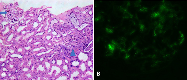
Histological examination of the renal biopsy specimen. (A) Light microscopy (H&E stain) showing two glomeruli, the left one is unremarkable (arrow) and the right one depicts segmental mesangial and endocapillary hypercellularity (arrowhead). (B) Immunofluorescence microscopy showing brightly immunofluorescent IgA deposits in the mesangium and focally along the capillary walls.