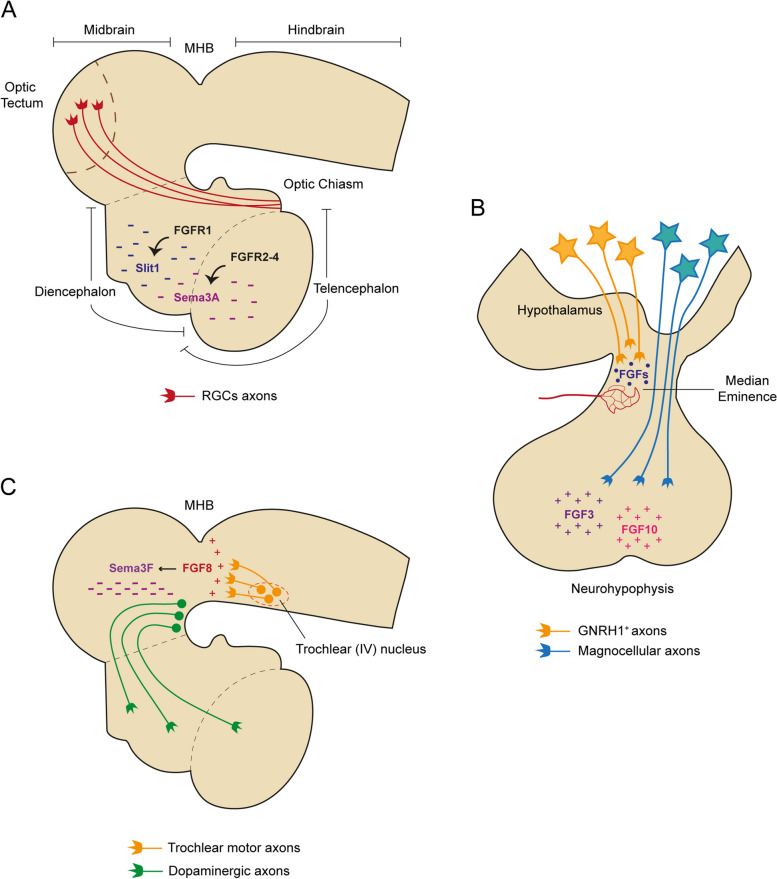
Fig. 4.
Role of FGF signaling in axon pathfinding. A Schematic diagram of the neural tube showing the regulation of the trajectory of RGCs axons by FGF signaling. In the Xenopus, slit1 and sema3A expression in the forebrain is positively regulated by FGFR1 and FGFR2-4 respectively. These two guidance molecules work together to repel RGCs axons out of the mid-diencephalon in the direction of the optic tectum (superior colliculus in mammals). B Schematic representation of the hypothalamus-hypophyseal system. During development, neurons that synthesize GNRH1 send their axons to the median eminence to ensure the release of GNRH1 into circulation to reach the adenohypophysis (not shown). FGFs emanating from the median eminence may act as chemoattractive cues, since the expression of a dominant negative form of FGFR1 in GNRH1 neurons compromises the targeting of their axons to this region. By contrast, magnocellular axons traverse the median eminence and reach the neurohypophysis where they release the peptides vasopressin and oxytocin into the general circulation. In the chick brain, FGF3 and FGF10 secreted by the neurohypophysis attract these hypothalamic neurosecretory axons towards this region. C Side view of the neural tube showing the effect of FGF8 signaling in axon pathfinding. FGF8 produced by the isthmic organizer at the MHB attracts trochlear motor axons as they extend from cell bodies in the anterior hindbrain. This MHB-derived FGF8 also regulates the growth of midbrain dopaminergic axons by inducing the expression of sema3F in the midbrain. This repulsive cue guides dopaminergic axons rostrally towards their diencephalic and telencephalic targets. GNRH1, Gonadotropin-releasing hormone; MHB, Midbrain-hindbrain boundary; RGCs, Retinal ganglion cells

