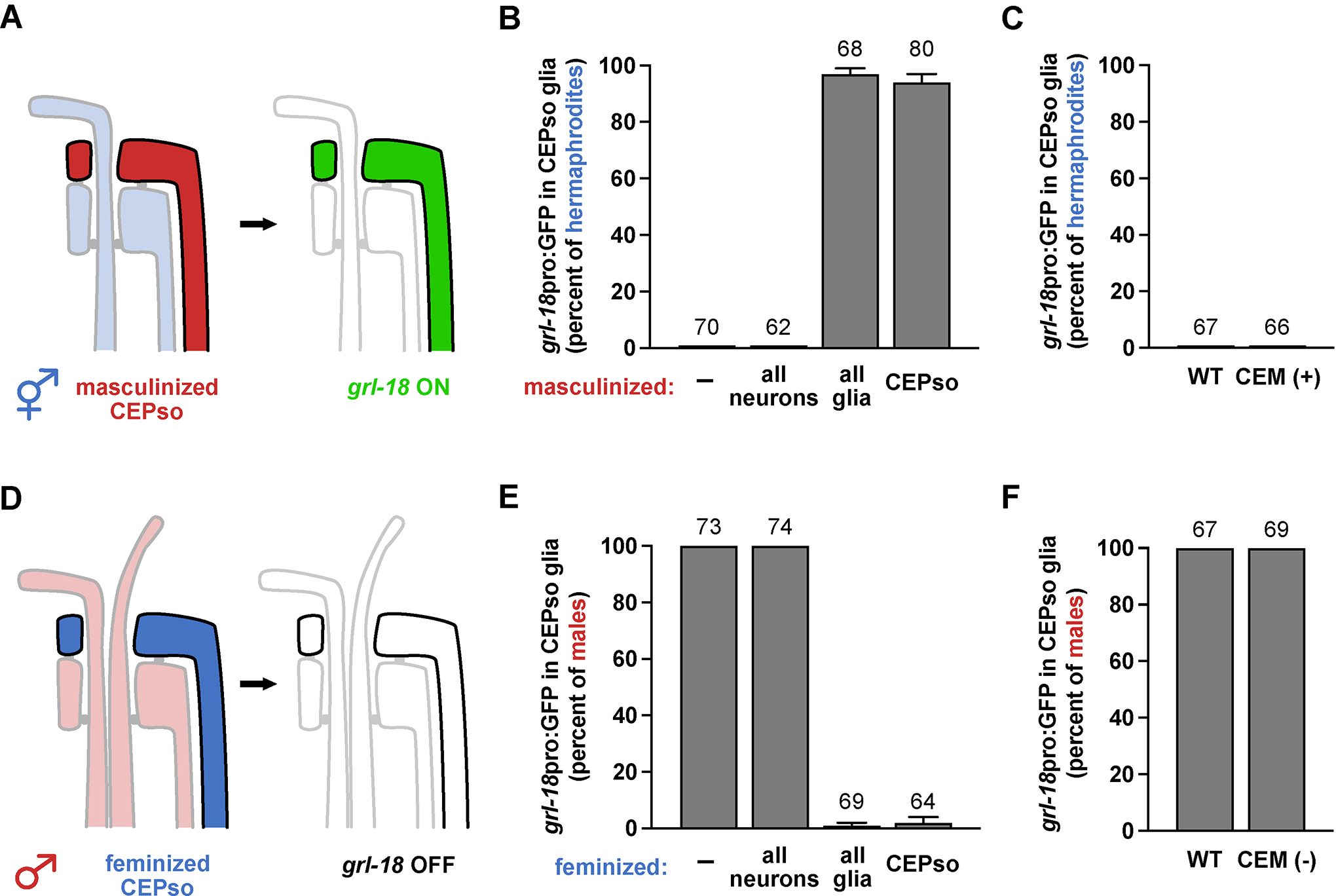
Figure 2. The sex-specific switch in gene expression is controlled cell autonomously in glia and does not depend on male neurons.

(A-B, D-E) Neurons or glia were (A) masculinized in hermaphrodites or (D) feminized in males via mis-expression of fem-3 or tra-2(IC), respectively, using cell-type-specific promoters (all neurons, rab-3; all glia, mir-228; CEPso glia, col-56, see Figure S2). Fraction of 1-day adult (B) hermaphrodites or (E) males that express grl-18pro:GFP in CEPso glia is shown. (C, F) To test if CEM neurons are sufficient and/or necessary for the switch, grl-18pro:GFP expression was evaluated in (C) ceh-30(n3714gf) hermaphrodites in which CEM neurons inappropriately survive (CEM+), and (F) in ceh-30(n4289lf) males in which CEM neurons inappropriately undergo apoptosis (CEM−)27. Fraction of 1-day adult (C) CEM+ hermaphrodites and (F) CEM− males that express grl-18pro:GFP in CEPso glia. Sample sizes are indicated above the bars of each graph. Error bars, SEM.
